
A) B) c) GEL O-ring GEL Air chamber NMR tube Funnel Figure6.Anisotropic environment created by strained gels.(A)3-mm gel,contained in a4.5-mm inner diameter Shigemi sample cell. prior to compression by the plunger.(B)After compression by the plunger.(C)Device for achieving radial compression,resulting in axial stretching of the gel
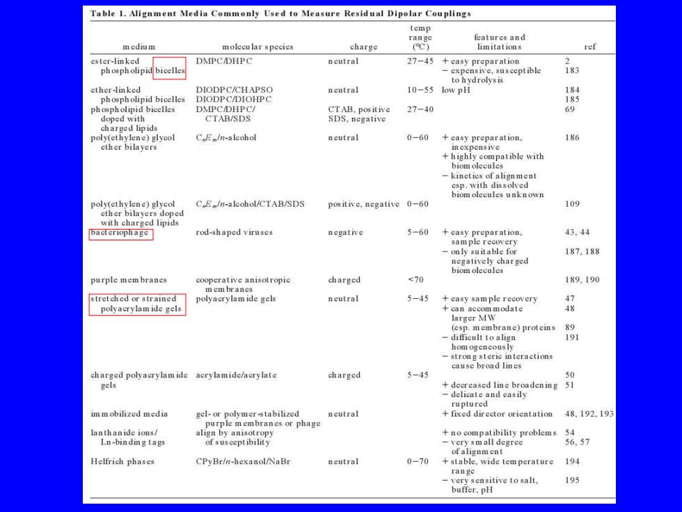
Table 1.Alignment Media Commonly Used to Measure Residual Dipolar Couplings temp features and medium molecu lar species charge (C) limitations ref ester-linked DMPC/DHPC neutral 27-45 +easy preparation phospholipid bicelles -expensive.susceptible 183 to hydrol小ysis ether-linked DIODPC/CHAPSO ncutral 10-55 low pH 184 phospholipid bicelles DIODPC/DIOHPC 185 pp CTAB.positive 27-40 69 CTAB/SDS SDS.negative charged lipids poly(ethylene)glycol C Em/n-aleohol neutral 0-60 +casy preparation, 186 ether bilayers inexpensive +highly compatible with biom olec kineties of alignment esp.with dissolved biom olecules unknown poly(ethylene)glycol C E/n-aleohol/CTAB/SDS positive.negative 0-60 109 ether bilayers doped with charged lipids rod-shaped viruses 5-60 +a 43.44 -only suitable for 187,188 negatively charged biomolecules purple mem branes cooperative anisotropic charged <70 189.190 mem brancs stretched or strained polyacrylam ide gels ncutral 5-45 +easy sample recovery polyacrylamide gels +nNmodae 4极 (esp.membrane)proteins 89 191 interactions charged polyacrylam ide acrylamide/acrylate charged 5-45 50 gels +decreased line broadening 51 -delicate and casily ruptured im m obilized media neutral +fixed director orientation 48.192,19 purple membranes or phage lanthanide ions/ align by anisotropy no compatibility problems 54 Ln-binding tags of susceptibility -very small degree 6.57 ofalignment Helfrich phases CPyBr/n-hexanol/NaBr neutral 0-70 +stable,wide tem perature 194 「ange very sensitive to salt, 195 buffer.pH
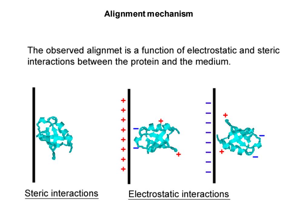
Alignment mechanism The observed alignmet is a function of electrostatic and steric interactions between the protein and the medium. ++ -- + Steric interactions Electrostatic interactions
Alignment mechanism
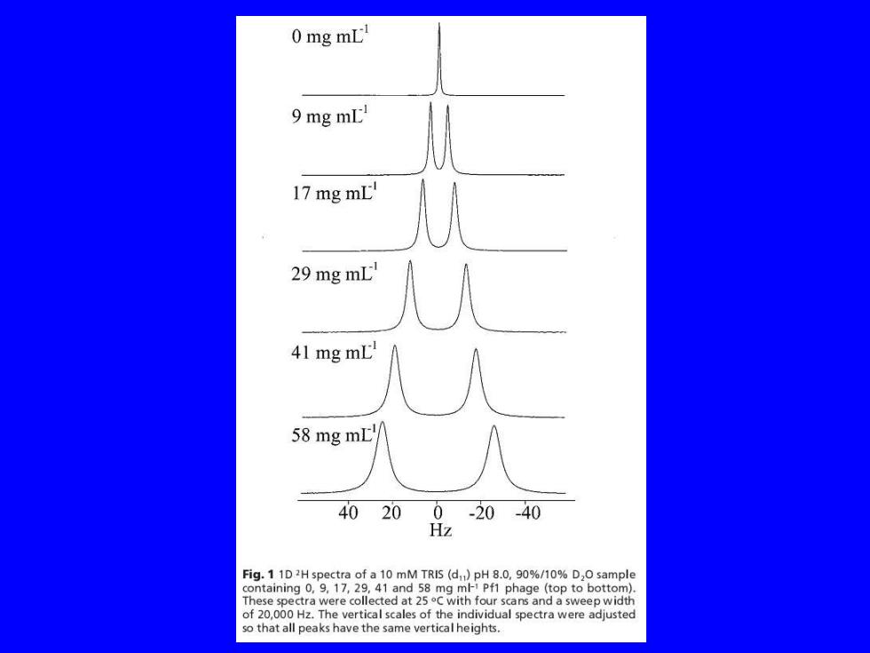
0 mg mL 9 mg mL 17 mg mL! 29 mg mL 41 mg mL 58 mg mLt 40 200-20-40 Hz Fig.1 1D2H spectra of a 10 mM TRIS (d)pH 8.0,90%/10%D,O sample containing 0,9.17,29,41 and 58 mg ml Pf1 phage (top to bottom). These spectra were collected at 25C with four scans and a sweep width of 20,000 Hz.The vertical scales of the individual spectra were adjusted so that all peaks have the same vertical heights
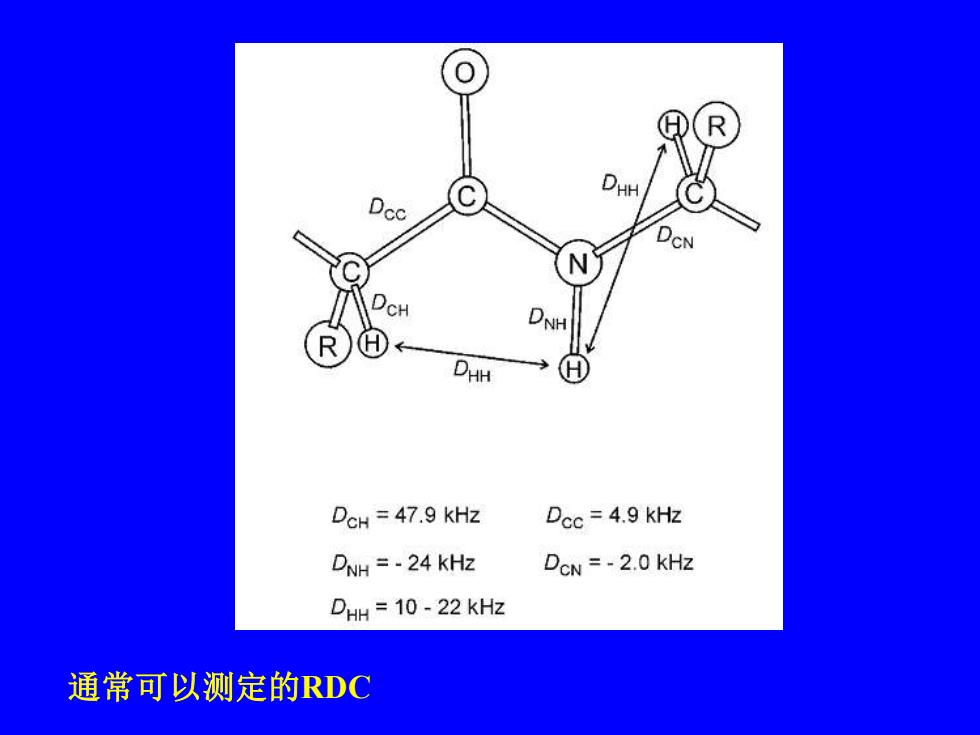
CN H DHH DCH 47.9 kHz Dcc=4.9 KHz DNH =-24 KHz DoN =-2.0 KHz DHH 10-22 kHz 通常可以测定的RDC
通常可以测定的RDC