
English Reading Materials Chapter 19:Agents Used in Cardiac Arrhythmias INTRODUCTION Cardiac arrhythmias are a frequent problem in clinical practice,occurring in up to 25%of patients treated with digitalis,50%of anesthetized patients,and over 80%of patients with acute myocardial infarction.Arrhythmias may require treatment because rhythms that are too rapid,too slow,or asynchronous can reduce cardiac output.Some arrhythmias can precipitate more serious or even lethal rhythm disturbances eg, early premature ventricular depolarizations can precipitate ventricular fibrillation.In such patients,antiarrhythmic drugs may be lifesaving.On the other hand,the hazards of antiarrhythmic drugs and in particular the fact that they can precipitate lethal arrhythmias in some patients has led to a reevaluation of their relative risks and benefits.In general,treatment of asymptomatic or minimally symptomatic arrhythmias should be avoided for this reason. Arrhythmias can be treated with the drugs discussed in this chapter and with nonpharmacologic therapies such as pacemakers,cardioversion,catheter ablation,and surgery.This chapter describes the pharmacology of drugs that suppress arrhythmias by a direct action on the cardiac cell membrane.Other modes of therapy are discussed briefly(see Box,The Nonpharmacologic Therapy of Cardiac Arrhythmias) THE NONPHARMACOLOGIC THERAPY OF CARDIAC ARRHYTHMIAS It was recognized over 100 years ago that reentry in simple in vitro models (eg,rings of conducting tissues)was permanently interrupted by transecting the reentry circuit. This concept is now applied in cardiac arrhythmias with defined anatomic pathways eg,atrioventricular reentry using accessory pathways,atrioventricular node reentry,atrial flutter,and some forms of ventricular tachycardia by treatment with radiofrequency catheter ablation.Recent studies have shown that paroxysmal and persistent atrial fibrillation may arise from one of the pulmonary veins.Both forms of atrial fibrillation can be cured by electrically isolating the pulmonary veins by radiofrequency catheter ablation or during concomitant cardiac surgery. Another form of nonpharmacologic therapy is the implantable cardioverter-defibrillator (ICD),a device that can automatically detect and treat potentially fatal arrhythmias such as ventricular fibrillation.ICDs are now widely used in patients who have been resuscitated from such arrhythmias,and several trials have shown that ICD treatment reduces mortality in patients with coronary artery 1
1 English Reading Materials Chapter 19: Agents Used in Cardiac Arrhythmias INTRODUCTION Cardiac arrhythmias are a frequent problem in clinical practice, occurring in up to 25% of patients treated with digitalis, 50% of anesthetized patients, and over 80% of patients with acute myocardial infarction. Arrhythmias may require treatment because rhythms that are too rapid, too slow, or asynchronous can reduce cardiac output. Some arrhythmias can precipitate more serious or even lethal rhythm disturbances eg, early premature ventricular depolarizations can precipitate ventricular fibrillation. In such patients, antiarrhythmic drugs may be lifesaving. On the other hand, the hazards of antiarrhythmic drugs and in particular the fact that they can precipitate lethal arrhythmias in some patients has led to a reevaluation of their relative risks and benefits. In general, treatment of asymptomatic or minimally symptomatic arrhythmias should be avoided for this reason. Arrhythmias can be treated with the drugs discussed in this chapter and with nonpharmacologic therapies such as pacemakers, cardioversion, catheter ablation, and surgery. This chapter describes the pharmacology of drugs that suppress arrhythmias by a direct action on the cardiac cell membrane. Other modes of therapy are discussed briefly (see Box, The Nonpharmacologic Therapy of Cardiac Arrhythmias). THE NONPHARMACOLOGIC THERAPY OF CARDIAC ARRHYTHMIAS It was recognized over 100 years ago that reentry in simple in vitro models (eg, rings of conducting tissues) was permanently interrupted by transecting the reentry circuit. This concept is now applied in cardiac arrhythmias with defined anatomic pathways eg, atrioventricular reentry using accessory pathways, atrioventricular node reentry, atrial flutter, and some forms of ventricular tachycardia by treatment with radiofrequency catheter ablation. Recent studies have shown that paroxysmal and persistent atrial fibrillation may arise from one of the pulmonary veins. Both forms of atrial fibrillation can be cured by electrically isolating the pulmonary veins by radiofrequency catheter ablation or during concomitant cardiac surgery. Another form of nonpharmacologic therapy is the implantable cardioverter-defibrillator (ICD), a device that can automatically detect and treat potentially fatal arrhythmias such as ventricular fibrillation. ICDs are now widely used in patients who have been resuscitated from such arrhythmias, and several trials have shown that ICD treatment reduces mortality in patients with coronary artery
disease who have an ejection fraction 30%and in patients with class 2 or 3 heart failure and no prior history of arrhythmias.The increasing use of nonpharmacologic antiarrhythmic therapies reflects both advances in the relevant technologies and an increasing appreciation of the dangers of long-term therapy with currently available drugs. ELECTROPHYSIOLOGY OF NORMAL CARDIAC RHYTHM Introduction The electrical impulse that triggers a normal cardiac contraction originates at regular intervals in the sinoatrial node(Figure 1),usually at a frequency of 60-100 beats per minute.This impulse spreads rapidly through the atria and enters the atrioventricular node,which is normally the only conduction pathway between the atria and ventricles. Conduction through the atrioventricular node is slow,requiring about 0.15 s.(This delay provides time for atrial contraction to propel blood into the ventricles.)The impulse then propagates over the His-Purkinje system and invades all parts of the ventricles,beginning with the endocardial surface near the apex and ending with the epicardial surface at the base of the heart.Ventricular activation is complete in less than 0.1 s;therefore,contraction of all of the ventricular muscle is normally synchronous and hemodynamically effective. Arrhythmias consist of cardiac depolarizations that deviate from the above description in one or more aspects ie,there is an abnormality in the site oforigin of the impulse,its rate or regularity,or its conduction. 2
2 disease who have an ejection fraction 30% and in patients with class 2 or 3 heart failure and no prior history of arrhythmias. The increasing use of nonpharmacologic antiarrhythmic therapies reflects both advances in the relevant technologies and an increasing appreciation of the dangers of long-term therapy with currently available drugs. ELECTROPHYSIOLOGY OF NORMAL CARDIAC RHYTHM Introduction The electrical impulse that triggers a normal cardiac contraction originates at regular intervals in the sinoatrial node (Figure 1), usually at a frequency of 60-100 beats per minute. This impulse spreads rapidly through the atria and enters the atrioventricular node, which is normally the only conduction pathway between the atria and ventricles. Conduction through the atrioventricular node is slow, requiring about 0.15 s. (This delay provides time for atrial contraction to propel blood into the ventricles.) The impulse then propagates over the His-Purkinje system and invades all parts of the ventricles, beginning with the endocardial surface near the apex and ending with the epicardial surface at the base of the heart. Ventricular activation is complete in less than 0.1 s; therefore, contraction of all of the ventricular muscle is normally synchronous and hemodynamically effective. Arrhythmias consist of cardiac depolarizations that deviate from the above description in one or more aspects ie, there is an abnormality in the site of origin of the impulse, its rate or regularity, or its conduction

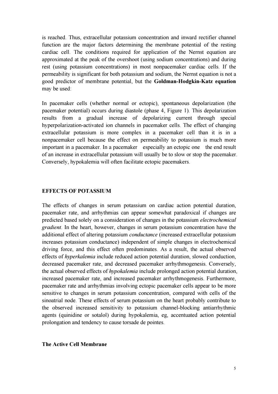
Superior vena cava Phase 0 SA node Atrium AV node Overshoot 0 Phase 0 mV Purkinje -100 Tricuspid Resting potentia valve Mitral valve Action potential phases Ventricle 0:Upstroke 1:Early-fast repolarization 2:Plateau 3:Repolarization 4:Diastole ECG Q S PR- QT 200ms Figure 1.Schematic representation of the heart and normal cardiac electrical activity (intracellular recordings from areas indicated and ECG).Sinoatrial node, atrioventricular node,and Purkinje cells display pacemaker activity (phase 4 depolarization).The ECG is the body surface manifestation of the depolarization and repolarization waves of the heart.The P wave is generated by atrial depolarization, the QRS by ventricular muscle depolarization,and the T wave by ventricular repolarization.Thus,the PR interval is a measure of conduction time from atrium to ventricle,and the QRS duration indicates the time required for all of the ventricular cells to be activated (ie,the intraventricular conduction time).The QT interval reflects the duration of the ventricular action potential. Ionic Basis of Membrane Electrical Activity The transmembrane potential of cardiac cells is determined by the concentrations of several ions chiefly sodium (Na),potassium (K),calcium (Ca2),and chloride (Cl)on either side of the membrane and the permeability of the membrane to each ion.These water-soluble ions are unable to freely diffuse across the lipid cell membrane in response to their electrical and concentration gradients;they require aqueous channels(specific pore-forming proteins)for such diffusion.Thus,ions move across cell membranes in response to their gradients only at specific times during the 3
3 Figure 1. Schematic representation of the heart and normal cardiac electrical activity (intracellular recordings from areas indicated and ECG). Sinoatrial node, atrioventricular node, and Purkinje cells display pacemaker activity (phase 4 depolarization). The ECG is the body surface manifestation of the depolarization and repolarization waves of the heart. The P wave is generated by atrial depolarization, the QRS by ventricular muscle depolarization, and the T wave by ventricular repolarization. Thus, the PR interval is a measure of conduction time from atrium to ventricle, and the QRS duration indicates the time required for all of the ventricular cells to be activated (ie, the intraventricular conduction time). The QT interval reflects the duration of the ventricular action potential. Ionic Basis of Membrane Electrical Activity The transmembrane potential of cardiac cells is determined by the concentrations of several ions chiefly sodium (Na+ ), potassium (K+ ), calcium (Ca2+), and chloride (Cl- ) on either side of the membrane and the permeability of the membrane to each ion. These water-soluble ions are unable to freely diffuse across the lipid cell membrane in response to their electrical and concentration gradients; they require aqueous channels (specific pore-forming proteins) for such diffusion. Thus, ions move across cell membranes in response to their gradients only at specific times during the

cardiac cycle when these ion channels are open.The movements of the ions produce currents that form the basis of the cardiac action potential.Individual channels are relatively ion-specific,and the flux of ions through them is thought to be controlled by "gates"(probably flexible peptide chains or energy barriers).Each type of channel has its own type of gate(sodium,calcium,and some potassium channels are each thought to have two types of gates),and each type of gate is opened and closed by specific transmembrane voltage,ionic,or metabolic conditions. At rest,most cells are not significantly permeable to sodium,but at the start of each action potential,they become quite permeable (see below).In electrophysiologic terms,the conductance of the fast sodium channel suddenly increases in response to a depolarizing stimulus.Similarly,calcium enters and potassium leaves the cell with each action potential.Therefore,in addition to ion channels,the cell must have mechanisms to maintain stable transmembrane ionic conditions by establishing and maintaining ion gradients.The most important of these active mechanisms is the sodium pump,Na/K ATPase.This pump and other active ion carriers contribute indirectly to the transmembrane potential by maintaining the gradients necessary for diffusion through channels.In addition,some pumps and exchangers produce net current flow(eg,by exchanging three Na"for two K*ions)and hence are termed "electrogenic." When the cardiac cell membrane becomes permeable to a specific ion (ie when the channels selective for that ion are open),movement of that ion across the cell membrane is determined by Ohm's law:current voltage resistance,or current voltage conductance.Conductance is determined by the properties of the individual ion channel protein.The voltage term is the difference between the actual membrane potential and the reversal potential for that ion(the membrane potential at which no current would flow even if channels were open).For example,in the case of sodium in a cardiac cell at rest,there is a substantial concentration gradient (140 mmol/L Na"outside;10-15 mmol/L Na"inside)and an electrical gradient (0 mV outside:-90 mV inside)that would drive Na'into cells.Sodium does not enter the cell at rest because sodium channels are closed;when sodium channels open,the very large influx of Na'ions accounts for phase 0 depolarization.The situation for K*ions in the resting cardiac cell is quite different.Here,the concentration gradient (140 mmol/L inside;4 mmol/L outside)would drive the ion out of the cell,but the electrical gradient would drive it in;that is,the inward gradient is in equilibrium with the outward gradient.In fact,certain potassium channels("inward rectifier"channels) are open in the resting cell,but little current flows through them because of this balance.The equilibrium,or reversal potential,for ions is determined by the Nernst equation: where Ce and Ci are the extracellular and intracellular concentrations,respectively, multiplied by their activity coefficients.Note that raising extracellular potassium makes Ek less negative.When this occurs,the membrane depolarizes until the new Ek 4
4 cardiac cycle when these ion channels are open. The movements of the ions produce currents that form the basis of the cardiac action potential. Individual channels are relatively ion-specific, and the flux of ions through them is thought to be controlled by "gates" (probably flexible peptide chains or energy barriers). Each type of channel has its own type of gate (sodium, calcium, and some potassium channels are each thought to have two types of gates), and each type of gate is opened and closed by specific transmembrane voltage, ionic, or metabolic conditions. At rest, most cells are not significantly permeable to sodium, but at the start of each action potential, they become quite permeable (see below). In electrophysiologic terms, the conductance of the fast sodium channel suddenly increases in response to a depolarizing stimulus. Similarly, calcium enters and potassium leaves the cell with each action potential. Therefore, in addition to ion channels, the cell must have mechanisms to maintain stable transmembrane ionic conditions by establishing and maintaining ion gradients. The most important of these active mechanisms is the sodium pump, Na+ /K+ ATPase. This pump and other active ion carriers contribute indirectly to the transmembrane potential by maintaining the gradients necessary for diffusion through channels. In addition, some pumps and exchangers produce net current flow (eg, by exchanging three Na+ for two K+ ions) and hence are termed "electrogenic." When the cardiac cell membrane becomes permeable to a specific ion (ie when the channels selective for that ion are open), movement of that ion across the cell membrane is determined by Ohm's law: current = voltage resistance, or current = voltage conductance. Conductance is determined by the properties of the individual ion channel protein. The voltage term is the difference between the actual membrane potential and the reversal potential for that ion (the membrane potential at which no current would flow even if channels were open). For example, in the case of sodium in a cardiac cell at rest, there is a substantial concentration gradient (140 mmol/L Na+ outside; 10-15 mmol/L Na+ inside) and an electrical gradient (0 mV outside; -90 mV inside) that would drive Na+ into cells. Sodium does not enter the cell at rest because sodium channels are closed; when sodium channels open, the very large influx of Na+ ions accounts for phase 0 depolarization. The situation for K+ ions in the resting cardiac cell is quite different. Here, the concentration gradient (140 mmol/L inside; 4 mmol/L outside) would drive the ion out of the cell, but the electrical gradient would drive it in; that is, the inward gradient is in equilibrium with the outward gradient. In fact, certain potassium channels ("inward rectifier" channels) are open in the resting cell, but little current flows through them because of this balance. The equilibrium, or reversal potential, for ions is determined by the Nernst equation: where Ce and Ci are the extracellular and intracellular concentrations, respectively, multiplied by their activity coefficients. Note that raising extracellular potassium makes EK less negative. When this occurs, the membrane depolarizes until the new EK
is reached.Thus,extracellular potassium concentration and inward rectifier channel function are the major factors determining the membrane potential of the resting cardiac cell.The conditions required for application of the Nernst equation are approximated at the peak of the overshoot (using sodium concentrations)and during rest (using potassium concentrations)in most nonpacemaker cardiac cells.If the permeability is significant for both potassium and sodium,the Nernst equation is not a good predictor of membrane potential,but the Goldman-Hodgkin-Katz equation may be used: In pacemaker cells (whether normal or ectopic),spontaneous depolarization (the pacemaker potential)occurs during diastole (phase 4,Figure 1).This depolarization results from a gradual increase of depolarizing current through special hyperpolarization-activated ion channels in pacemaker cells.The effect of changing extracellular potassium is more complex in a pacemaker cell than it is in a nonpacemaker cell because the effect on permeability to potassium is much more important in a pacemaker.In a pacemaker especially an ectopic one the end result of an increase in extracellular potassium will usually be to slow or stop the pacemaker. Conversely,hypokalemia will often facilitate ectopic pacemakers. EFFECTS OF POTASSIUM The effects of changes in serum potassium on cardiac action potential duration, pacemaker rate,and arrhythmias can appear somewhat paradoxical if changes are predicted based solely on a consideration of changes in the potassium electrochemical gradient.In the heart,however,changes in serum potassium concentration have the additional effect of altering potassium conductance (increased extracellular potassium increases potassium conductance)independent of simple changes in electrochemical driving force,and this effect often predominates.As a result,the actual observed effects of hyperkalemia include reduced action potential duration,slowed conduction, decreased pacemaker rate,and decreased pacemaker arrhythmogenesis.Conversely, the actual observed effects of hypokalemia include prolonged action potential duration, increased pacemaker rate,and increased pacemaker arrhythmogenesis.Furthermore, pacemaker rate and arrhythmias involving ectopic pacemaker cells appear to be more sensitive to changes in serum potassium concentration,compared with cells of the sinoatrial node.These effects of serum potassium on the heart probably contribute to the observed increased sensitivity to potassium channel-blocking antiarrhythmic agents (quinidine or sotalol)during hypokalemia,eg,accentuated action potential prolongation and tendency to cause torsade de pointes. The Active Cell Membrane 5
5 is reached. Thus, extracellular potassium concentration and inward rectifier channel function are the major factors determining the membrane potential of the resting cardiac cell. The conditions required for application of the Nernst equation are approximated at the peak of the overshoot (using sodium concentrations) and during rest (using potassium concentrations) in most nonpacemaker cardiac cells. If the permeability is significant for both potassium and sodium, the Nernst equation is not a good predictor of membrane potential, but the Goldman-Hodgkin-Katz equation may be used: In pacemaker cells (whether normal or ectopic), spontaneous depolarization (the pacemaker potential) occurs during diastole (phase 4, Figure 1). This depolarization results from a gradual increase of depolarizing current through special hyperpolarization-activated ion channels in pacemaker cells. The effect of changing extracellular potassium is more complex in a pacemaker cell than it is in a nonpacemaker cell because the effect on permeability to potassium is much more important in a pacemaker. In a pacemaker especially an ectopic one the end result of an increase in extracellular potassium will usually be to slow or stop the pacemaker. Conversely, hypokalemia will often facilitate ectopic pacemakers. EFFECTS OF POTASSIUM The effects of changes in serum potassium on cardiac action potential duration, pacemaker rate, and arrhythmias can appear somewhat paradoxical if changes are predicted based solely on a consideration of changes in the potassium electrochemical gradient. In the heart, however, changes in serum potassium concentration have the additional effect of altering potassium conductance (increased extracellular potassium increases potassium conductance) independent of simple changes in electrochemical driving force, and this effect often predominates. As a result, the actual observed effects of hyperkalemia include reduced action potential duration, slowed conduction, decreased pacemaker rate, and decreased pacemaker arrhythmogenesis. Conversely, the actual observed effects of hypokalemia include prolonged action potential duration, increased pacemaker rate, and increased pacemaker arrhythmogenesis. Furthermore, pacemaker rate and arrhythmias involving ectopic pacemaker cells appear to be more sensitive to changes in serum potassium concentration, compared with cells of the sinoatrial node. These effects of serum potassium on the heart probably contribute to the observed increased sensitivity to potassium channel-blocking antiarrhythmic agents (quinidine or sotalol) during hypokalemia, eg, accentuated action potential prolongation and tendency to cause torsade de pointes. The Active Cell Membrane