
Plaque formation I-Fatty streak Monocyte Oxidized DL Foam cell Macrophage Figure 10 Monocytes penetrate the intima and are transformed into macrophages and eventually cholesterol-rich foam cells.These activated macrophages scavenge and ingest oxidized 图9-2主动脉粥样硬化 low-density lipoprotein(LDL)in the subendothelial space.The progressive accumulation of lipids 主动脉内膜表面可见隆起的脂纹,纤维斑块(ntra-and extracelluar)forms the fatty streak. Second Clinical Medical College of Southern Medical University
Second Clinical Medical College of Southern Medical University
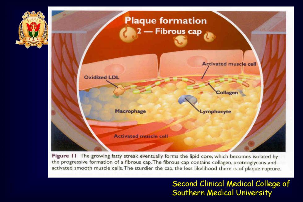
Plaque formation ★★ 2-Fibrous cap Activated muscle cell Oxidized LDL Collagen Macrophage Lymphocyte Activated muscle cell Figure II The growing fatty streak eventually forms the lipid core,which becomes isolated by the progressive formation of a fibrous cap.The fibrous cap contains collagen,proteoglycans and activated smooth muscle cells.The sturdier the cap,the less likelihood there is of plaque rupture. Second Clinical Medical College of Southern Medical University
Second Clinical Medical College of Southern Medical University

Plaque formation 3-Lipid core Lipid accumulation Foam cells Free radicals Cell death Figure 12 Further lipid accumulation in the lipid core results in cell death(apoptosis) Second Clinical Medical College of Southern Medical University
Second Clinical Medical College of Southern Medical University

动脉粥样硬化视频 SAMPLE USE ONLY nucleus e2011 Second Clinical Medical College of Southern Medical University
Second Clinical Medical College of Southern Medical University 动脉粥样硬化视频

4 最大冠脉流量 冠脉内径减少 3 达到正常75% 2 一80%以上时, 安静时冠脉流童 会引起冠状动 脉血流量明显 0 20 40 60 减少。 80 100 冠脉狭窄程度(%) 冠脉血流自我 调节功能。 图2一】冠脉狭窄程度与冠脉流量的关系 (引自Marcus ML) Southern Medical University
Second Clinical Medical College of Southern Medical University •冠脉内径减少 达到正常75% 一80%以上时, 会引起冠状动 脉血流量明显 减少。 •冠脉血流自我 调节功能