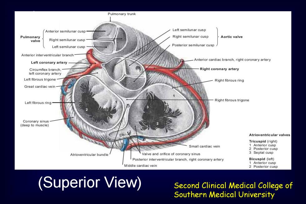
Pulmonary trunk Anterior semilunar cusp Left semilunar cusp Pulmonary Right semilunar cusp Aortic valve valve Right semilunar cusp Left semilunar cusp Posterior semilunar cusp Anterior interventricular branch Anterior cardiac branch.right coronary artery Left coronary artery Circumflex branch Right coronary artery left coronary artery Left fibrous trigone- Right fibrous ring Great cardiac vein Left fibrous ring Right fibrous trigone Coronary sinus (deep to muscle】 Atrioventricular valves Tricuspid(right) Small cardiac vein Anterior cusp 2 Posterior cusp Atrioventricular bundle Valve and orifice of coronary sinus 3 Septal cusp Posterior interventricular branch,right coronary artery Bicuspid (left) 1 Anterior cusp Middle cardiac vein 2 Posterior cusp (Superior View) Second Clinical Medical College of Southern Medical University
Second Clinical Medical College of Southern Medical University (Superior View)
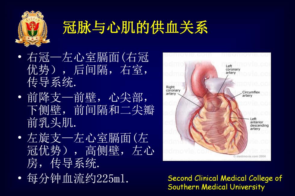
冠脉与心肌的供血关系 右冠一左心室膈面(右冠 优势),后间隔,右室, Left coronary artery 传导系统 。 前降支一前壁,心尖部, 下侧壁,前间隔和二尖 前乳头肌. descending artery 左旋支一左心室膈面(左 冠优势),高侧壁,左心 房,传导系统 每分钟血流约225m1. Second Clinical Medical College of Southern Medical University
Second Clinical Medical College of Southern Medical University 冠脉与心肌的供血关系 • 右冠—左心室膈面(右冠 优势),后间隔,右室, 传导系统. • 前降支—前壁,心尖部, 下侧壁,前间隔和二尖瓣 前乳头肌. • 左旋支—左心室膈面(左 冠优势),高侧壁,左心 房,传导系统. • 每分钟血流约225ml
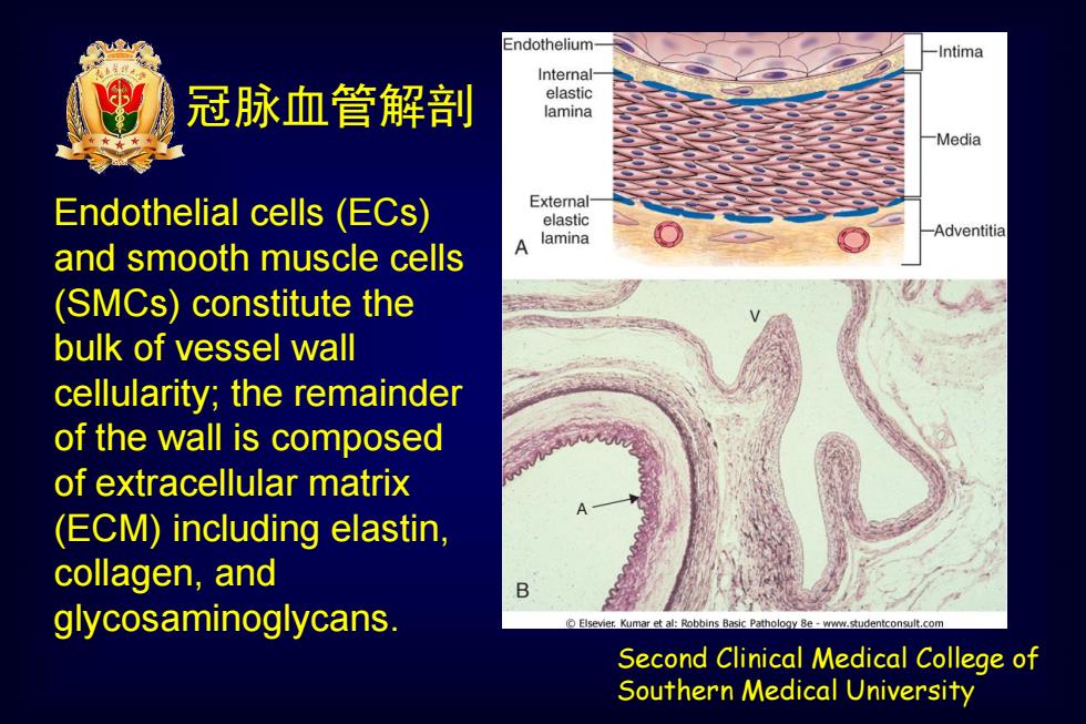
Endothelium- -Intima Internal- 冠脉血管解剖 elastic lamina -Media Endothelial cells (ECs) External- elastic and smooth muscle cells A lamina Adventitia (SMCs)constitute the bulk of vessel wall cellularity;the remainder of the wall is composed of extracellular matrix (ECM)including elastin, collagen,and B glycosaminoglycans. Elsevier.Kumar e et al:Robbins Basic Pathology 8e-www.stu ntconsult.con Second Clinical Medical College of Southern Medical University
Second Clinical Medical College of Southern Medical University Endothelial cells (ECs) and smooth muscle cells (SMCs) constitute the bulk of vessel wall cellularity; the remainder of the wall is composed of extracellular matrix (ECM) including elastin, collagen, and glycosaminoglycans. 冠脉血管解剖

血管阻力 血容量分布 47% Small arteries and arterioles 64% Veins 19% 9%/ 27% Arteries 7% Lung Capillaries 7% Heart Veins (diastole) 5% 8% 7% Capillaries Distribution of Small arteries Large vascular resistance and arterioles arteries in the systemic circulation Volume distribution Second Clinical Medical College of Southern Medical University
Second Clinical Medical College of Southern Medical University 血容量分布 血管阻力
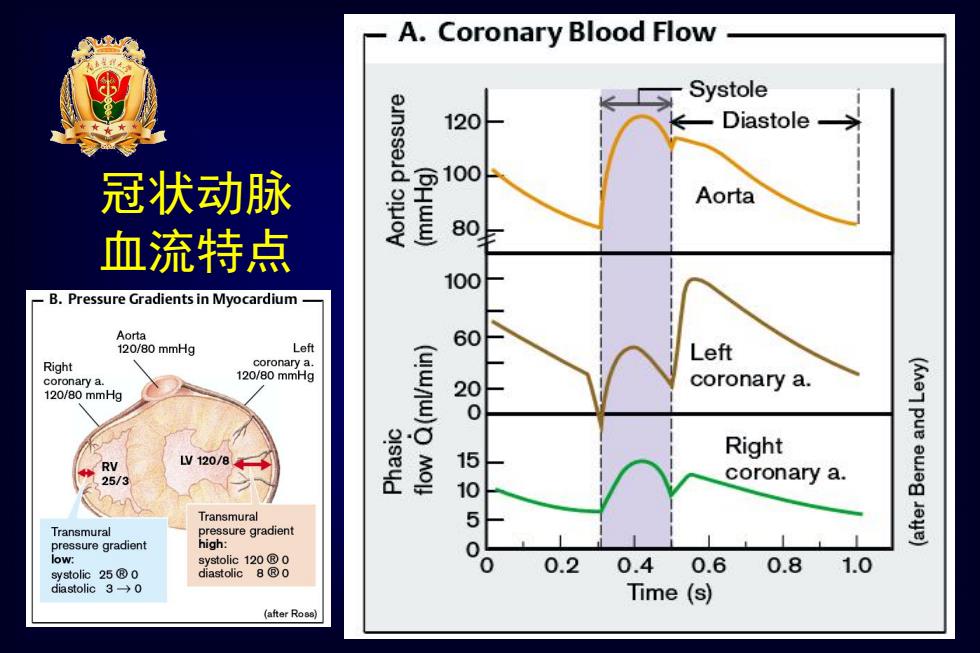
A. Coronary Blood Flow Systole o 120 Diastole 冠状动脉 Aorta 血流特点 ww 80 100 B.Pressure Gradients in Myocardium- Aorta 120/80mmHg Left 60 Left Right coronary a. coronary a. 120/80mmHg 120/80mmHg (uwnw) 28 coronary a. (Kne7 pue Right RV L120/84 MO 15 25/3 coronary a. 10 eueg Transmural Transmural pressure gradient eye) pressure gradient high: 50 low: systolic120®0 systolic25®0 diastolic8®o 0.2 0.4 0.6 0.8 1.0 diastolic3→0 Time (s) (after Ross)
Second Clinical Medical College of Southern Medical University 冠状动脉 血流特点