
conjunctiva cillary body 1.Fibrous layer: Providing shape and resistance rectus t Cornea: optlc axis- -visual axis Covering ant.1/6,transparent,rich N. vitreous humor Endothelial cells do not regenerate. Corneal transplantation. choroid lamina cribosa Section human ey Sclera: nacula lutea optic nerve sheath Covering the post.5/6. Providing attachment for muscles. Scleral venous sinus: an irregular circular canal beneath the junction of cornea and sclera
1. Fibrous layer: Providing shape and resistance Cornea: Covering ant. 1/6, transparent, rich N. Endothelial cells do not regenerate. Corneal transplantation. Sclera: Covering the post. 5/6. Providing attachment for muscles. Scleral venous sinus: an irregular circular canal beneath the junction of cornea and sclera

2.Vascular layer Iris-pupil,controlling the light into the eye Ciliary body Ciliary muscle -control the lens thickness Zonular fibers Ciliary processes-~80,producing AH Choroid-supplying nutrition absorbing the disperse light Circular muscles Pupil Radial muscles contract contract Bright light Normal light Dim light
2. Vascular layer Iris – pupil, controlling the light into the eye Ciliary body - Ciliary muscle - control the lens thickness Zonular fibers Ciliary processes - ~80, producing AH Choroid – supplying nutrition absorbing the disperse light

2.Vascula Connective tissue Cilliary body. Iris-pupil,con Ciliary body Ciliary muscl Zonular fibers Ciliary proces Cilliary Choroid-supp processes absorbing Zonule fibers Circular muscles contract Lens Bright light
2. Vascular layer Iris – pupil, controlling the light into the eye Ciliary body - Ciliary muscle - control the lens thickness Zonular fibers Ciliary processes - ~80, producing AH Choroid – supplying nutrition absorbing the disperse light
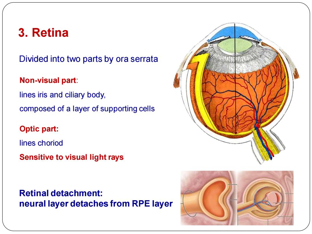
3.Retina Divided into two parts by ora serrata Non-visual part: lines iris and ciliary body, composed of a layer of supporting cells Optic part: lines choriod Sensitive to visual light rays Retinal detachment: neural layer detaches from RPE layer
3. Retina Non-visual part: lines iris and ciliary body, composed of a layer of supporting cells Optic part: lines choriod Sensitive to visual light rays Divided into two parts by ora serrata Retinal detachment: neural layer detaches from RPE layer

Light Vitreous body Inner limiting membrane VB 10 Optic nerve fibres 9 Ganglion cell layer 8 Inner plexiform layer Inner nuclear layer Outer plexiform layer 5 Cell bodies of rods and cones Outer limiting membrane 3 Photoreceptor layer Pigment cells Choroid