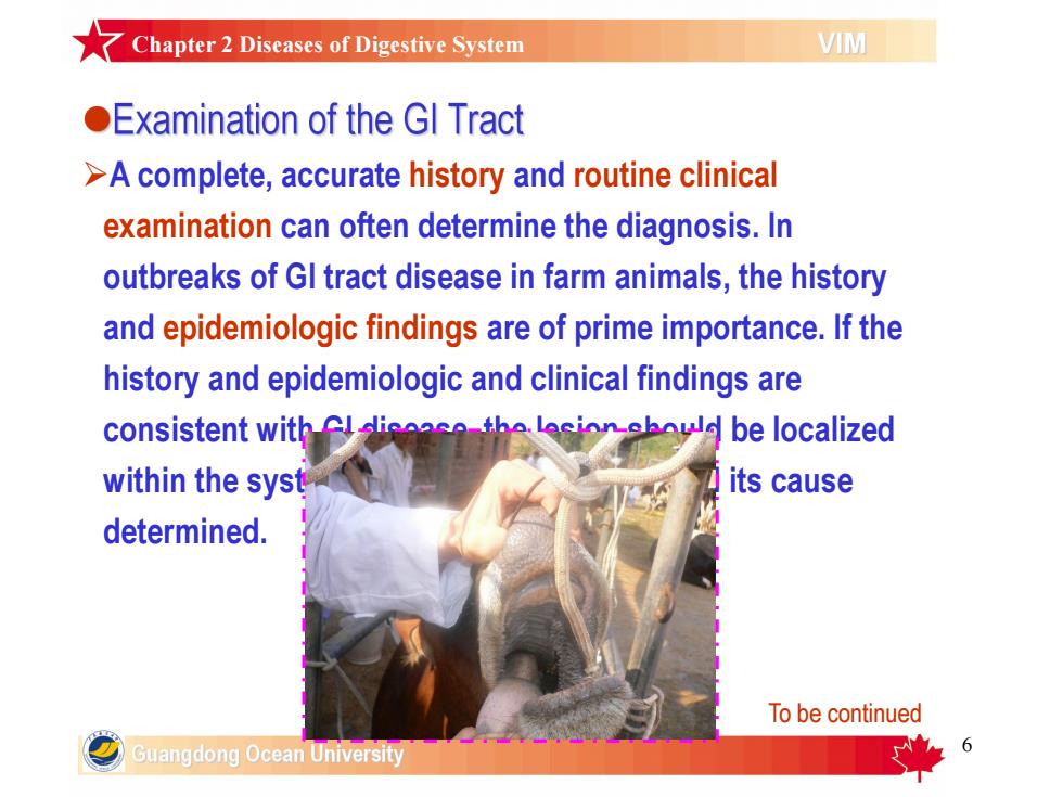
Chapter2Diseases of DigestiveSystemVIMExamination of the GI Tract>Acomplete,accuratehistoryandroutineclinicalexamination can often determine the diagnosis.Inoutbreaks of Gl tract disease in farm animals, the historyand epidemiologic findings are of prime importance.If thehistoryand epidemiologic and clinical findings aretha lacion ehould be localizedconsistent with Cl-iTitscausewithin the syst拉determined.To be continuedGuangdong Ocean University
6 ØA complete, accurate history and routine clinical examination can often determine the diagnosis. In outbreaks of GI tract disease in farm animals, the history and epidemiologic findings are of prime importance. If the history and epidemiologic and clinical findings are consistent with GI disease, the lesion should be localized within the system, and the type of lesion and its cause determined. To be continued Chapter 2 Diseases of Digestive System
★Chapter2Diseases of Digestive SystemVIMExaminationoftheGl Tract1) Visual inspection of the oral cavity,and of thecontourof theabdomen for distention orcontraction;2)Palpation through theabdominal wall or per rectum to evaluate shape, size,and position ofabdominal viscera;3)Abdominal percussion todetect“pings,"which suggestgas-filledviscera;4)Auscultation todetermine the intensity,frequency,andduration of GI movements,as wellas fluid-splashing sounds associated with fluid-filled stomachs and intestines and fluidrushing sounds associated with diarrheal disease;5)Succussion猛摇torevealfluid-splashingsounds;6) Ballottement浮球感to evaluatedensityand sizeofabdominal organsbytheirmovementawayfrom and backto the abdominal wall;7)Grossexamination肉眼观察offecestoassessbulk,consistency软硬度,color,andpresenceofmucus,blood,orundigestedfood particles.To be continuedGuangdong Ocean University
7 1) Visual inspection of the oral cavity, and of the contour of the abdomen for distention or contraction; 2) Palpation through the abdominal wall or per rectum to evaluate shape, size, and position of abdominal viscera; 3) Abdominal percussion to detect “pings, ” which suggest gas-filled viscera; 4) Auscultation to determine the intensity, frequency, and duration of GI movements, as well as fluid-splashing sounds associated with fluid-filled stomachs and intestines and fluid- rushing sounds associated with diarrheal disease; 5) Succussion猛摇 to reveal fluid-splashing sounds; 6) Ballottement浮球感 to evaluate density and size of abdominal organs by their movement away from and back to the abdominal wall; 7) Gross examination 肉眼观察of feces to assess bulk, consistency软硬度, color, and presence of mucus, blood, or undigested food particles. To be continued Chapter 2 Diseases of Digestive System
7Chapter2 Diseases of Digestive SystemVIMExaminationoftheGlTractThe following may be useful (or necessary):Bacterial culture and virus isolation;2)Endoscopyto visualizethemucosal surface oftheesophagus,stomachduo'denum+二指肠,colon,andrectum;3)Abdominocentesis腹腔穿刺术tocollectfluidfromdistendedvisceraorfrom the peritoneal cavity for examination;4)Radiography (contrast)to diagnose obstructive disease;5)Abdominal ultrasonography to detect abdominal masses,intussusceptions肠套叠,and mesen'teric lymphadenopathy in smallanimals, and to investigate abdominal disorders in horses and cows;6)Biopsy活检(endoscopic,laparoscopic,surgical)toobtainsamplesformicroscopicexamination(samples of intestines and liver are useful indiagnosing chronic enteritis and liver disease);7Testsfor digestionandabsorptiontoestimate and differentiatemalabsorption and maldigestion.Guangdong Ocean University
8 Ø The following may be useful (or necessary): 1) Bacterial culture and virus isolation; 2) Endoscopy to visualize the mucosal surface of the esophagus, stomach, duo’denum十二指肠, colon, and rectum; 3) Abdominocentesis腹腔穿刺术 to collect fluid from distended viscera or from the peritoneal cavity for examination; 4) Radiography (contrast) to diagnose obstructive disease; 5) Abdominal ultrasonography to detect abdominal masses, intussusceptions肠套叠, and mesen’teric lymphadenopathy in small animals, and to investigate abdominal disorders in horses and cows; 6) Biopsy活检 (endoscopic, laparoscopic, surgical) to obtain samples for microscopic examination (samples of intestines and liver are useful in diagnosing chronic enteritis and liver disease); 7) Tests for digestion and absorption to estimate and differentiate malabsorption and maldigestion. Chapter 2 Diseases of Digestive System
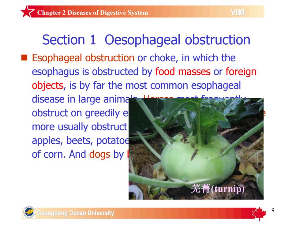
ZChapter2Diseases of Digestive SystemVIMSection 1 Oesophageal obstructionEsophageal obstructionorchoke,inwhichtheesophagus is obstructed by food masses or foreignobjects,isbyfarthemostcommonesophagealdisease in large animalobstruct on greedily emore usually obstructapples,beets,potatoeof corn.And dogs by芜菁(turnip)GuangdongOcean University
9 Section 1 Oesophageal obstruction n Esophageal obstruction or choke, in which the esophagus is obstructed by food masses or foreign objects, is by far the most common esophageal disease in large animals. Horses most frequently obstruct on greedily eaten dried grains or hay. Cattle more usually obstruct on a single solid object, eg, apples, beets, potatoes, turnips, corn stalks, or ears of corn. And dogs by bones (when dog is scared). 芜菁(turnip) Chapter 2 Diseases of Digestive System
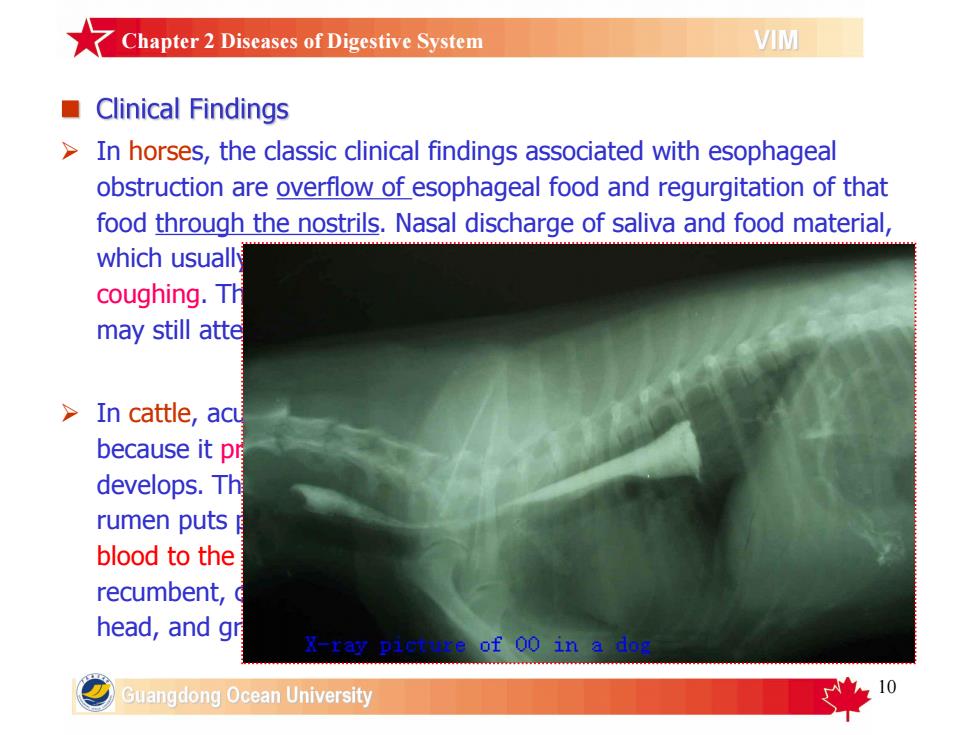
7 Chapter2 Diseases of Digestive SystemVIMClinical Findings In horses, the classic clinical findings associated with esophagealobstruction are overflow of esophageal food and regurgitation of thatfood through the nostrils. Nasal discharge of saliva and food material,which usuallycoughing. Thmay still atte> In cattle,acubecause it prdevelops. Thrumen puts blood to therecumbent, head, and grof00inGuangdong Ocean University
10 Ø In horses, the classic clinical findings associated with esophageal obstruction are overflow of esophageal food and regurgitation of that food through the nostrils. Nasal discharge of saliva and food material, which usually also spills from the pharynx into the airway, induces coughing. The horse is anxious and may stretch and arch its neck but may still attempt to continue to either eat or drink. Ø In cattle, acute and complete esophageal obstruction is an emergency because it prohibits the eructation of ruminal gases, and free-gas bloat develops. This in turn may result in asphyxia(窒息) as the expanding rumen puts pressure on the diaphragm(膜膈) and reduces return of blood to the heart. The cow may be bloated and in distress or recumbent, or there may be protrusion of the tongue, extension of the head, and grinding of the teeth with excess salivation. Chapter 2 Diseases of Digestive System