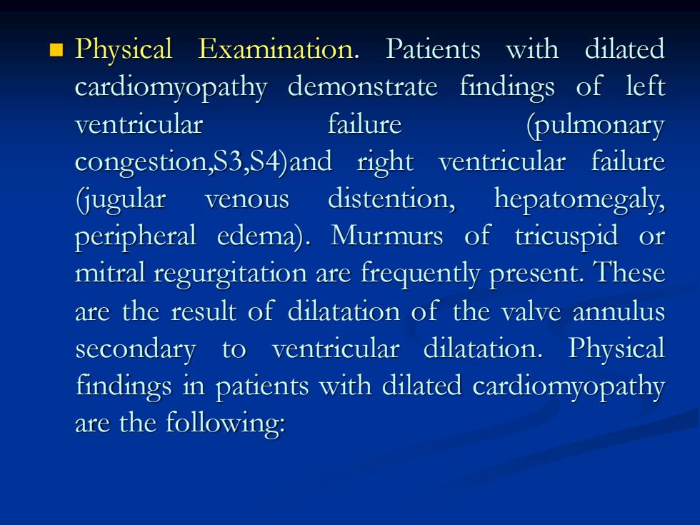
Physical Examination.Patients with dilated cardiomyopathy demonstrate findings of left ventricular failure (pulmonary congestion,S3,S4)and right ventricular failure (jugular venous distention,hepatomegaly, peripheral edema).Murmurs of tricuspid or mitral regurgitation are frequently present.These are the result of dilatation of the valve annulus secondary to ventricular dilatation.Physical findings in patients with dilated cardiomyopathy are the following:
◼ Physical Examination. Patients with dilated cardiomyopathy demonstrate findings of left ventricular failure (pulmonary congestion,S3,S4)and right ventricular failure (jugular venous distention, hepatomegaly, peripheral edema). Murmurs of tricuspid or mitral regurgitation are frequently present. These are the result of dilatation of the valve annulus secondary to ventricular dilatation. Physical findings in patients with dilated cardiomyopathy are the following:
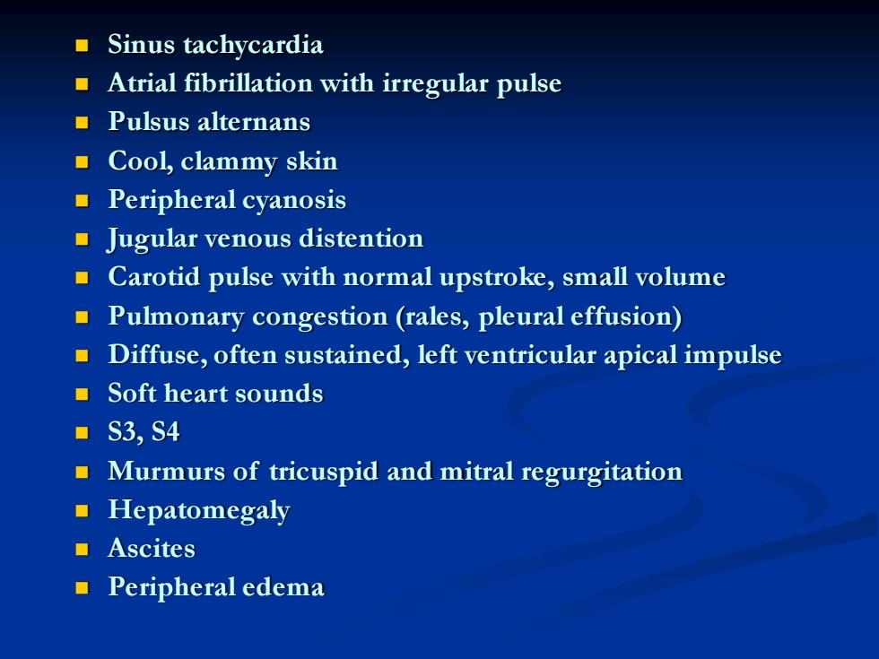
■Sinus tachycardia Atrial fibrillation with irregular pulse ■Pulsus alternans ■Cool,clammy skin ■Peripheral cyanosis Jugular venous distention Carotid pulse with normal upstroke,small volume Pulmonary congestion(rales,pleural effusion) Diffuse,often sustained,left ventricular apical impulse ■Soft heart sounds ·S3,S4 Murmurs of tricuspid and mitral regurgitation ■Hepatomegaly ■Ascites ■Peripheral edema
◼ Sinus tachycardia ◼ Atrial fibrillation with irregular pulse ◼ Pulsus alternans ◼ Cool, clammy skin ◼ Peripheral cyanosis ◼ Jugular venous distention ◼ Carotid pulse with normal upstroke, small volume ◼ Pulmonary congestion (rales, pleural effusion) ◼ Diffuse, often sustained, left ventricular apical impulse ◼ Soft heart sounds ◼ S3, S4 ◼ Murmurs of tricuspid and mitral regurgitation ◼ Hepatomegaly ◼ Ascites ◼ Peripheral edema

ECG.Electrocardiographic findings in patients with dilated cardiomyopathy include low-voltage QRS complexes,conduction defects,and atrial or ventricular (or both)arrhythmias.Holter monitoring or exercise testing also may disclose malignant ventricular arrhythmias such as ventricular tachycardia.The ECG findings in dilated cardiomyopathy are summarized in Table 2
◼ ECG. Electrocardiographic findings in patients with dilated cardiomyopathy include low-voltage QRS complexes, conduction defects, and atrial or ventricular (or both) arrhythmias. Holter monitoring or exercise testing also may disclose malignant ventricular arrhythmias such as ventricular tachycardia. The ECG findings in dilated cardiomyopathy are summarized in Table 2

Chest x-ray examination.Left ventricular and often left atrial enlargement will be apparent on the chest x-ray films of patients with dilated cardiomyopathy.Occasionally,right ventricular enlargement is also seen.Massive cardiomegaly may be observed on the chest film,and it is sometimes difficult to distinguish dilated cardiomyopathy from large pericardial effusion by chest film alone.Signs of pulmonary venous hypertension are also frequently discernble in the chest roentgenograms of patients with dilated cardiomyopathy.Roentgenographic findings in patients with dilated cardiomyopathy are summarized in Table 2
◼ Chest x-ray examination. Left ventricular and often left atrial enlargement will be apparent on the chest x-ray films of patients with dilated cardiomyopathy. Occasionally, right ventricular enlargement is also seen. Massive cardiomegaly may be observed on the chest film, and it is sometimes difficult to distinguish dilated cardiomyopathy from large pericardial effusion by chest film alone. Signs of pulmonary venous hypertension are also frequently discernble in the chest roentgenograms of patients with dilated cardiomyopathy. Roentgenographic findings in patients with dilated cardiomyopathy are summarized in Table 2

Table 2.Eletrocardiographic and roentgenographic findings in dilated cardiomyopathy Eletrcardiogaphic findings ■Atrial fibrillation Ventricular arrhythmias Low-voltage QRS complexes Nonspecific T wave changes(flattening,inversion) ■ Conduction defects(e.g.,LBBB) Left ventricular hypertrophy Poor R wave progression in precordial leads mimicking anterior MI Chest x-ray findings Left ventricular enlargement Left atrial enlargement Right ventricular enlargement(less common) Signs of pulmonary congestion(pulmonary vascular redistribution, interstitial edema)
Table 2 . Eletrocardiographic and roentgenographic findings in dilated cardiomyopathy Eletrcardiogaphic findings ◼ Atrial fibrillation ◼ Ventricular arrhythmias ◼ Low-voltage QRS complexes ◼ Nonspecific T wave changes(flattening, inversion) ◼ Conduction defects(e.g., LBBB) ◼ Left ventricular hypertrophy ◼ Poor R wave progression in precordial leads mimicking anterior MI Chest x-ray findings ◼ Left ventricular enlargement ◼ Left atrial enlargement ◼ Right ventricular enlargement (less common) ◼ Signs of pulmonary congestion (pulmonary vascular redistribution, interstitial edema)