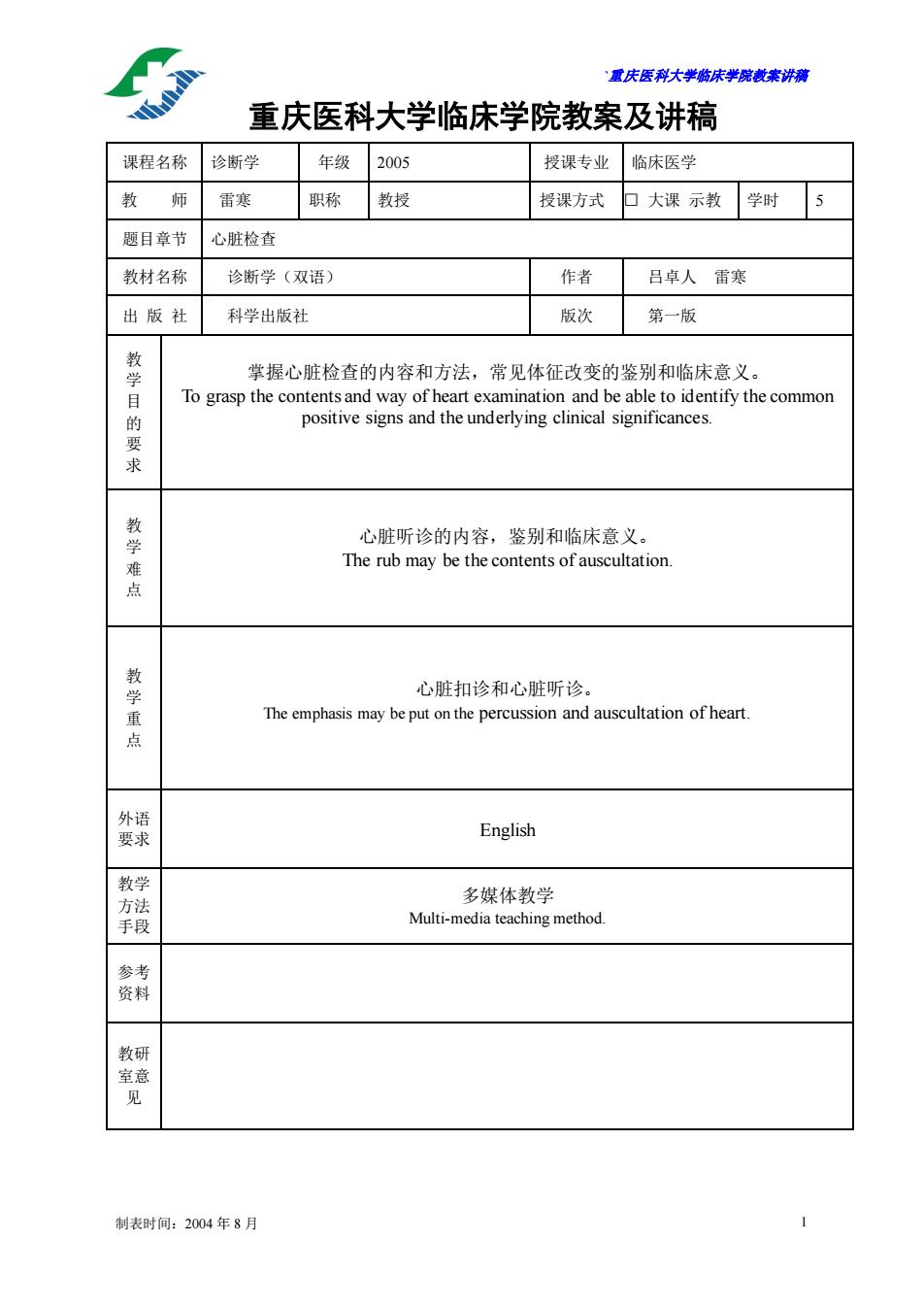
置庆医科大学脑床学院讲满 重庆医科大学临床学院教案及讲稿 课程名称诊断学 年级2005 授课专业临床医学 教师雷寒 职称教授 授课方式口大课示教学时5 题目章节心脏检查 教材名称诊断学(双语) 作者吕卓人雷寒 出版社 科学出版社 版次 第一版 掌握心脏检查的内容和方法,常见体征改变的鉴别和临床意义。 求 心脏听诊的内容,鉴别和临床意义。 The r rub may be the contents of auscultation 点 教学重点 心脏扣诊和心脏听诊。 The emphasis may be put on the percussion and auscultation of heart. 父 English 多媒体教学 Multi-edia teaching method 花 制表时间:2004年8月
`重庆医科大学临床学院教案讲稿 制表时间:2004 年 8 月 1 重庆医科大学临床学院教案及讲稿 课程名称 诊断学 年级 2005 授课专业 临床医学 教 师 雷寒 职称 教授 授课方式 □ 大课 示教 学时 5 题目章节 心脏检查 教材名称 诊断学(双语) 作者 吕卓人 雷寒 出 版 社 科学出版社 版次 第一版 教 学 目 的 要 求 掌握心脏检查的内容和方法,常见体征改变的鉴别和临床意义。 To grasp the contents and way of heart examination and be able to identify the common positive signs and the underlying clinical significances. 教 学 难 点 心脏听诊的内容,鉴别和临床意义。 The rub may be the contents of auscultation. 教 学 重 点 心脏扣诊和心脏听诊。 The emphasis may be put on the percussion and auscultation of heart. 外语 要求 English 教学 方法 手段 多媒体教学 Multi-media teaching method. 参考 资料 教研 室意 见
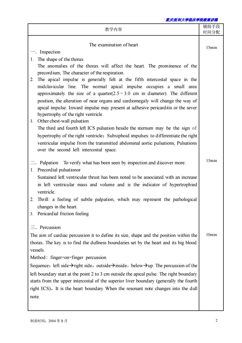
露庆医科大学临床半蕊裁案讲满 教学内容 辅助手段 时间分配 The examination of heart 15min Inspection 1.The shape of the thorax The anomalies of the thorax will affect the heart.The prominence of the precordium;The character of the respiration. 2 The apical impulse is generally fet at the fifth intercostal space in the midclavicular line.The normal apical impulse occupies a small area approximately the size of a quarter(2.5-3.0 cm in diameter).The different position,the alteration of near organs and cardiomegaly will change the way of apical impulse.Inward impulse may present at adhesive pericarditis or the sever The third and fourth left ICS pulsation beside the sterum may be the sign of hypertrophy of the right ventricle:Subxiphoid impulses:to differentiate the righ ventricular impulse from the transmitted abdominal aortic pulsations;Pulsations over the second left intercostal space Palpation To verify what has been seen by inspection and discover more. 15min 1.Precordial pulsationor Sustained left ventricular thrust has been noted to be associated with an increase in left ventricular mass and volume and is the indicator of hypertrophied ventricle 2 Thrill:a feeling of subtle palpation,which may represent the pathological changes in the heart. Pericardial friction feeling 三.Percussion The aim of cardiac percussion it to define its size,shape and the position within the 10min thorax.The key is to find the dullness boundaries set by the heart and its big blood vessels. Method:finger-on-finger percussion. Sequence:left sideright side,outside>inside,belowup.The percussion of the left boundary start at the point 2 to3 cm outside the apical pulse.The right boundary starts from the upper intercostal of the superior liver boundary (generally the fourth right ICS).It is the heart boundary When the resonant note changes into the dul note. 制表时间:2004年8月
`重庆医科大学临床学院教案讲稿 制表时间:2004 年 8 月 2 教学内容 辅助手段 时间分配 The examination of heart 一.Inspection 1. The shape of the thorax The anomalies of the thorax will affect the heart. The prominence of the precordium; The character of the respiration. 2. The apical impulse is generally felt at the fifth intercostal space in the midclavicular line. The normal apical impulse occupies a small area approximately the size of a quarter(2.5–3.0 cm in diameter). The different position, the alteration of near organs and cardiomegaly will change the way of apical impulse. Inward impulse may present at adhesive pericarditis or the sever hypertrophy of the right ventricle. 3. Other chest-wall pulsation The third and fourth left ICS pulsation beside the sternum may be the sign of hypertrophy of the right ventricle;Subxiphoid impulses: to differentiate the right ventricular impulse from the transmitted abdominal aortic pulsations; Pulsations over the second left intercostal space. 二.Palpation To verify what has been seen by inspection and discover more. 1. Precordial pulsationor Sustained left ventricular thrust has been noted to be associated with an increase in left ventricular mass and volume and is the indicator of hypertrophied ventricle. 2. Thrill: a feeling of subtle palpation, which may represent the pathological changes in the heart. 3. Pericardial friction feeling 三.Percussion The aim of cardiac percussion it to define its size, shape and the position within the thorax. The key is to find the dullness boundaries set by the heart and its big blood vessels. Method: finger-on-finger percussion. Sequence:left side→right side,outside→inside,below→up. The percussion of the left boundary start at the point 2 to 3 cm outside the apical pulse. The right boundary starts from the upper intercostal of the superior liver boundary (generally the fourth right ICS)。It is the heart boundary When the resonant note changes into the dull note. 15min 15min 10min
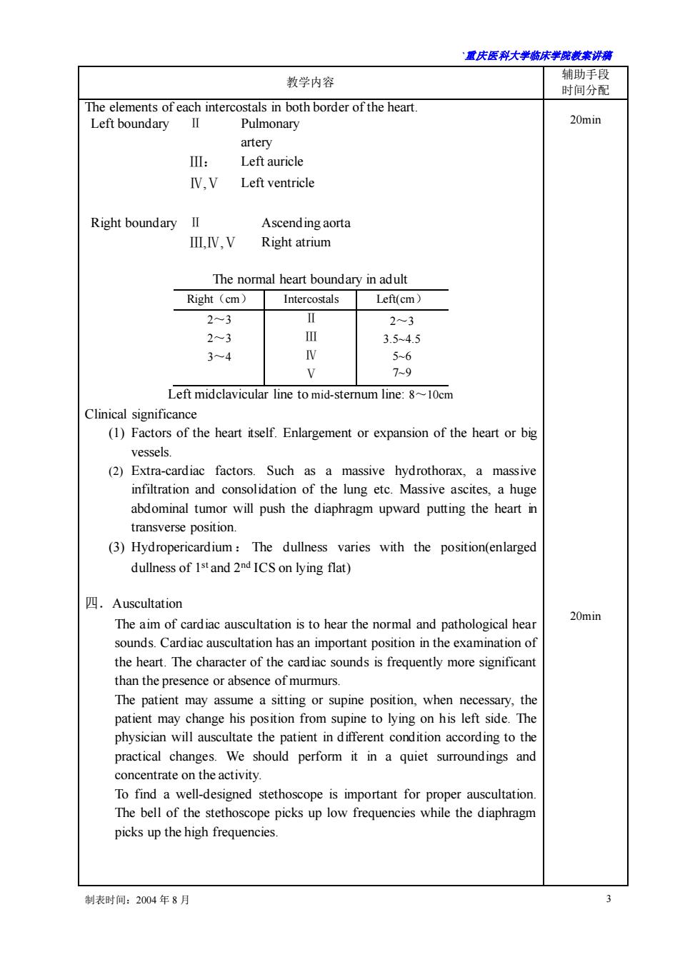
重庆医科大半床半院载未讲满 教学内容 辅助手段 时间分配 The elements of each intercostals in both border of the heart. Left boundaryⅡ Pulmonary 20min artery : Left auricle IV,V Left ventricle Right boundary II Ascending aorta M.IV.V Right atrium The normal heart boundary in adult Right(cm) Intercostals Left(cm) 2~3 2-3 3.54.5 56 9 Left midclavicular line to mid-sternum line:810cm Clinical significance (1)Factors of the heart itself.Enlargement or expansion of the heart or big vessels (2)Extra-cardiac factors. Such as a massive hydrothorax,a massive infiltration and consolidation of the lung etc.Massive ascites.a huge abdominal tumor will push the diaphragm upward putting the heart in transverse position. (3)Hydropericardium:The dullness varies with the position(enlarged dullness of 1s and 2ndICS on lying flat) 四.Auscultation The aim of cardiac auscultation is to hear the normal and pathological hea 20min sounds.Cardiac auscultation has an important position in the examination of the heart.The character of the cardiac sounds is frequently more significant than the presence or absence of murmurs. The patient may assume a sitting or supine position,when necessary,the patient may change his position from supine to lying on his left side.The physician will auscultate the patient in different condition according to the practical changes.We should perform it in a quiet surroundings and concentrate on the activity. To find a well-designed stethoscope is important for proper auscultation The bell of the stethoscope picks up low frequencies while the diaphragm picks up the high frequencies. 制表时间:2004年8月
`重庆医科大学临床学院教案讲稿 制表时间:2004 年 8 月 3 教学内容 辅助手段 时间分配 The elements of each intercostals in both border of the heart. Left boundary Ⅱ Pulmonary artery Ⅲ: Left auricle Ⅳ,Ⅴ Left ventricle Right boundary Ⅱ Ascending aorta Ⅲ,Ⅳ,Ⅴ Right atrium The normal heart boundary in adult Right(cm) Intercostals Left(cm) 2~3 2~3 3~4 Ⅱ Ⅲ Ⅳ Ⅴ 2~3 3.5~4.5 5~6 7~9 Left midclavicular line to mid-sternum line: 8~10cm Clinical significance (1) Factors of the heart itself. Enlargement or expansion of the heart or big vessels. (2) Extra-cardiac factors. Such as a massive hydrothorax, a massive infiltration and consolidation of the lung etc. Massive ascites, a huge abdominal tumor will push the diaphragm upward putting the heart in transverse position. (3) Hydropericardium : The dullness varies with the position(enlarged dullness of 1st and 2nd ICS on lying flat) 四.Auscultation The aim of cardiac auscultation is to hear the normal and pathological hear sounds. Cardiac auscultation has an important position in the examination of the heart. The character of the cardiac sounds is frequently more significant than the presence or absence of murmurs. The patient may assume a sitting or supine position, when necessary, the patient may change his position from supine to lying on his left side. The physician will auscultate the patient in different condition according to the practical changes. We should perform it in a quiet surroundings and concentrate on the activity. To find a well-designed stethoscope is important for proper auscultation. The bell of the stethoscope picks up low frequencies while the diaphragm picks up the high frequencies. 20min 20min
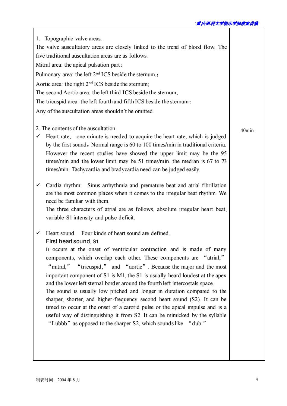
君庆医科大学临床半院表来讲满 1.Topographic valve areas The valve auscultatory areas are closely linked to the trend of blood flow.Th five traditional auscultation areas are as follows. Mitral area:the apical pulsation part: Pulmonary area:the left2nICS beside the stemum. Aortic area:the right 2 IC beside the stemum The second Aortic area:the left third ICS beside the sternum: The tricuspid area:the eft fourth and fifthICS beside the stemum: Any of the auscultation areas shouldn't be omitted. 2.The contents of the auscultation. 40min Heart rate:one minute is needed to acquire the heart rate,which is judged by the first sound.Normal range is 60 to 100 times/min in traditional criteria However the recent studies have showed the upper limit may be the 95 times/min and the lower limit may be 51 times/min.the median is 67 to 73 times/min.Tachycardia and bradycardia need can be judged easily. Cardia rhythm:Sinus arrhythmia and premature beat and atrial fibrillation are the most common places when it comes to the irregular beat rhythm.We need be familiar with them. The three characters of atrial are as follows,absolute irregular heart beat, variable SI intensity and pulse deficit. Heart sound.Four kinds of heart sound are defined. First heartsound,S1 It occurs at the onset of ventricular contraction and is made of man components,which overlap each other.These components are "atrial," “mitral,”“tricuspid,”and“aortic”.Because the major and the mos important component of S1 is M1,the S1 is usually heard loudest at the apex and the lower left stemal border around the fourth left intercostals space. The sound is usually low pitched and longer in duration compared to the sharper,shorter,and higher-frequency second heart sound (S2).It can be timed to occur at the onset of a carotid pulse or the apical impulse and is a useful way of distinguishng it from.It can be mimicked by the yllabe “Lubbb”as opposed to the sharper S2,which sounds like“dub.' 制表时间:2004年8月 4
`重庆医科大学临床学院教案讲稿 制表时间:2004 年 8 月 4 1. Topographic valve areas. The valve auscultatory areas are closely linked to the trend of blood flow. The five traditional auscultation areas are as follows. Mitral area: the apical pulsation part; Pulmonary area: the left 2nd ICS beside the sternum.; Aortic area: the right 2 nd ICS beside the sternum; The second Aortic area: the left third ICS beside the sternum; The tricuspid area: the left fourth and fifth ICS beside the sternum; Any of the auscultation areas shouldn’t be omitted. 2. The contents of the auscultation. ✓ Heart rate; one minute is needed to acquire the heart rate, which is judged by the first sound。Normal range is 60 to 100 times/min in traditional criteria. However the recent studies have showed the upper limit may be the 95 times/min and the lower limit may be 51 times/min. the median is 67 to 73 times/min. Tachycardia and bradycardia need can be judged easily. ✓ Cardia rhythm: Sinus arrhythmia and premature beat and atrial fibrillation are the most common places when it comes to the irregular beat rhythm. We need be familiar with them. The three characters of atrial are as follows, absolute irregular heart beat, variable S1 intensity and pulse deficit. ✓ Heart sound. Four kinds of heart sound are defined. First heart sound, S1 It occurs at the onset of ventricular contraction and is made of many components, which overlap each other. These components are “atrial,” “mitral,” “tricuspid,” and “aortic”. Because the major and the most important component of S1 is M1, the S1 is usually heard loudest at the apex and the lower left sternal border around the fourth left intercostals space. The sound is usually low pitched and longer in duration compared to the sharper, shorter, and higher-frequency second heart sound (S2). It can be timed to occur at the onset of a carotid pulse or the apical impulse and is a useful way of distinguishing it from S2. It can be mimicked by the syllable “Lubbb”as opposed to the sharper S2, which sounds like “dub.” 40min
置庆医科大学床半院未讲满 教学内容 Second heart sound,S2 The second heart sound occurs at the end of the ejection phase of systole.It is related to the closure of the semilunar valves.Since there are two semilunar valves,aortic and pulmonary,there are also two components for the 2 namely the aortic component(A2)and the pulmonary component (P2).The S is usually sharper,crisper,and shorter in duration compared to S1. The A2 component is normally heard over the true aortic area,which is the sash area (the area extending from the second right intercostal space at the sternal border to the apex).P2,on the other hand,is heard over the second and the third left intercostals spaces near the sternal border.Therefore,the splitting of the normal S2 is best appreciated at the second and third left intercosta space.Young people:P2>A2;Middle age man:P2=A2;Old age man:A2>P2 Third heart sound,S3: The early phase of the ventricular filling is characterized by sudden vigorous expansion associated with rapid inflow of blood.The peak of this filling period may be accompanied by a sound,which is termed the third heart sound,or S3 in children and in pregnant women. the S3 is a low-frequency sound or a thud similar to the sound caused by a small lead ball falling on a cushioned floor.It is obviously best audible over the apex area Fourth heart sound,S4 The atrium normally contracts at the end of diastole and gives an extra stretch to the ventricles.When the ventricular compliance is significantly reduced then this evokes a more vigorous contraction from the atrium,which forms the vibration.The sound is sometimes referred to as atrial gallop. Differentiate the Sland S2. The changes of heart sound and its clinical significance 40min (1)thenchanges of intensity of S1 and S2. S1 intensity depends on the degree of acceleration achieved by the contracting myofibrils at the time of mitral valve closure.Pressure rise (dP/dt)in the ventricle plays an important part.Increased atrial pressure will enhance the pressure rise (dP/dt),which may be obvious in mitral stenosis,incomplete atrial relaxation.Noncardiac factors such as body shape or chest wall deformities or the presence of pulmonary disease will functiontoo Severe mitral regurgitation will decrease the intensity of S1. 制表时间:2004年8月
`重庆医科大学临床学院教案讲稿 制表时间:2004 年 8 月 5 教学内容 辅助手段 时间分配 Second heart sound, S2: The second heart sound occurs at the end of the ejection phase of systole. It is related to the closure of the semilunar valves. Since there are two semilunar valves, aortic and pulmonary, there are also two components for the S2, namely the aortic component (A2) and the pulmonary component (P2). The S2 is usually sharper, crisper, and shorter in duration compared to S1. The A2 component is normally heard over the true aortic area, which is the sash area (the area extending from the second right intercostal space at the sternal border to the apex). P2, on the other hand, is heard over the second and the third left intercostals spaces near the sternal border. Therefore, the splitting of the normal S2 is best appreciated at the second and third left intercostal space. Young people:P2>A2; Middle age man: P2=A2; Old age man: A2>P2 Third heart sound, S3: The early phase of the ventricular filling is characterized by sudden vigorous expansion associated with rapid inflow of blood. The peak of this filling period may be accompanied by a sound, which is termed the third heart sound, or S3. This occurs in children and in pregnant women. the S3 is a low-frequency sound or a thud similar to the sound caused by a small lead ball falling on a cushioned floor. It is obviously best audible over the apex area Fourth heart sound, S4 The atrium normally contracts at the end of diastole and gives an extra stretch to the ventricles. When the ventricular compliance is significantly reduced, then this evokes a more vigorous contraction from the atrium, which forms the vibration. The sound is sometimes referred to as atrial gallop. Differentiate the S1and S2. The changes of heart sound and its clinical significance. (1) then changes of intensity of S1 and S2. S1 intensity depends on the degree of acceleration achieved by the contracting myofibrils at the time of mitral valve closure. Pressure rise (dP/dt) in the ventricle plays an important part. Increased atrial pressure will enhance the pressure rise (dP/dt) , which may be obvious in mitral stenosis, incomplete atrial relaxation. Noncardiac factors such as body shape or chest wall deformities or the presence of pulmonary disease will function too. Severe mitral regurgitation will decrease the intensity of S1. 40min