Exercise 2 Sample picture for Kirby-Bauer Antibiotic Susceptibility Tests Challenge:What is the genus of the following gram(-)coccus? Images: Streptocococcus pyogenes on blood agar Streptococcus agalactiae on blood agar file://CINDOWS/Desktop/clown/Microb Lab Web 2001/Exercise2/exercise2.htm (9of10)[8/3/0024:30:02 PM
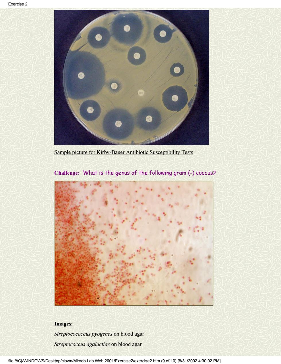
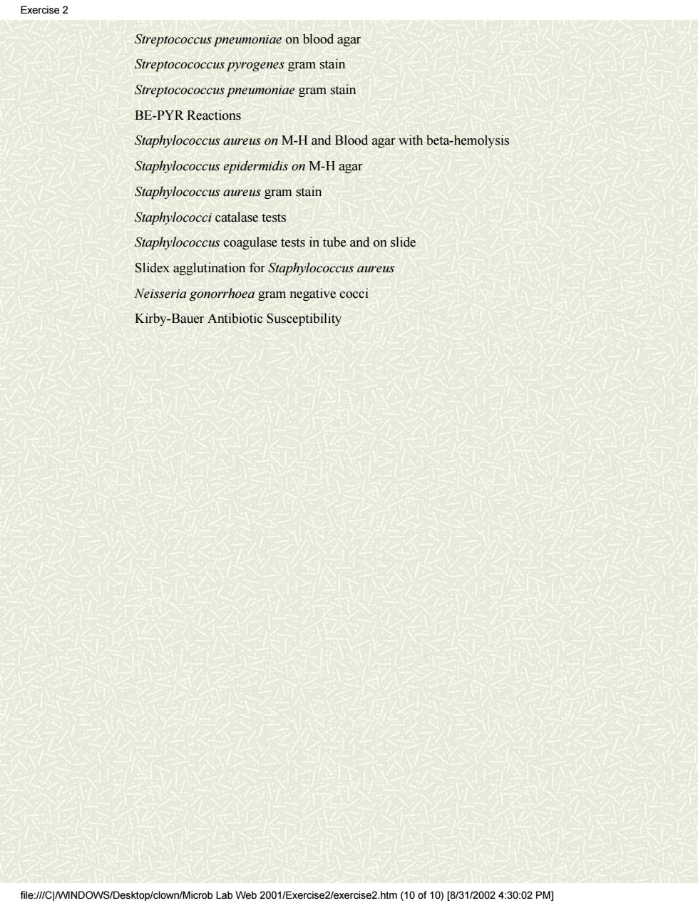
Sample picture for Kirby-Bauer Antibiotic Susceptibility Tests Challenge: What is the genus of the following gram (-) coccus? Images: Streptocococcus pyogenes on blood agar Streptococcus agalactiae on blood agar Exercise 2 file:///C|/WINDOWS/Desktop/clown/Microb Lab Web 2001/Exercise2/exercise2.htm (9 of 10) [8/31/2002 4:30:02 PM]
Exercise2 Streptococcus pneumoniae on blood agar Streptocococcus pyrogenes gram stain Streptocococcus pneumoniae gram stain BE-PYR Reactions Staphylococcus aureus on M-H and Blood agar with beta-hemolysis Staphylococcus epidermidis on M-H agar Staphylococcus aureus gram stain Staphylococci catalase tests Staphylococcus coagulase tests in tube and on slide Slidex agglutination for Staphylococcus aureus Neisseria gonorrhoea gram negative cocc Kirby-Bauer Antibiotic Susceptibility file:///C/WINDOWS/Desktop/clown/Microb Lab Web 2001/Exercise2/exercise2.htm(10 of 10)[8/31/2002 4:30:02 PM
Streptococcus pneumoniae on blood agar Streptocococcus pyrogenes gram stain Streptocococcus pneumoniae gram stain BE-PYR Reactions Staphylococcus aureus on M-H and Blood agar with beta-hemolysis Staphylococcus epidermidis on M-H agar Staphylococcus aureus gram stain Staphylococci catalase tests Staphylococcus coagulase tests in tube and on slide Slidex agglutination for Staphylococcus aureus Neisseria gonorrhoea gram negative cocci Kirby-Bauer Antibiotic Susceptibility Exercise 2 file:///C|/WINDOWS/Desktop/clown/Microb Lab Web 2001/Exercise2/exercise2.htm (10 of 10) [8/31/2002 4:30:02 PM]
Exercise 3 Cover Exercisel EXERCISE #3.GRAM-POSITIVE AND GRAM-NEGATIVE RODS Exercise2 FIRST SESSION Exercise3 FOCI: Identification Gram positive rods (Clostridium perfringens and Exercise4 Clostridium sporogenes)by Macroscopic and Microscopic Metabolic Exercise5 Characteristics. Identification Gram negative rods(Escherichia coli.Proteus Exercise6 mirabilis.Pseudomonas aeruginosa)by Macroscopic Microscopic and metabolic Characteristics Exercise7 Clinical case studies A.GRAM-POSITIVE Suggestions Characteristics of Clostridia All species of Clostridium are anaerobic spore-forming bacilli whose natural Coumter habitat is the soil and the intestinal tract of man and animals.In man,they cause gas g ene,tetanus,or botulism.In the first two diseases.cuts puncture w re conta with soil l or fec s harboring contaminated food. Clostridium perfringens is the most common species which causes gas ns have lethal d he nclude the properties in (le splits lecithin n mammalian nich has hemoly necrouizingets.hvalurondase whichs found in the substance that cements tissue cells together,collagenase which digests collagen on subcutaneous tissue and muscles,and DNase.The net result of such exotoxins is an invasive and spreading infection with death of surrounding tissue,crepitation,foul-smelling discharge,toxemia,shock,and ultimately death of the patient.As a general rule,gas gangrene is a mixed infection with proteolytic clostridia,various cocci,and gram-negative rods pre: ent In clostridial infections may result only fasciitis or cellulitis without gas gangrene Clostridium tetani forms a distinctive terminal spore and is identified by toxin neutralization tests.Open wounds are contaminated with spores or vegetative cells and the organisms grow in the devitalized tissue.Exotoxin(tetanus toxin)which is produced and secreted reaches the anterior horn cells of the spinal cord(probably via the peripheral nerves)where it diminishes inhibitory pses by blocking the al f ction of the inhibit smitter This ct of voluntar mu s which egin near the site of the infect (involv ment the jav ckaw) Tetanus neonatorium may result from infe tion ofth e umbilicus o newborn.To successfully cultivate anaerobes from clinical material. file://CWINDOWS/Desktop/clown/Microb Lab Web 2001/Exercise3/exercise3.htm(1 of7)[8/3/002 4:30:07 PM]
Cover Exercise1 Exercise2 Exercise3 Exercise4 Exercise5 Exercise6 Exercise7 Clinical case studies Suggestions Hit Counter EXERCISE #3. GRAM-POSITIVE AND GRAM-NEGATIVE RODS FIRST SESSION FOCI: Identification Gram positive rods (Clostridium perfringens and Clostridium sporogenes) by Macroscopic and Microscopic Metabolic Characteristics. Identification Gram negative rods (Escherichia coli, Proteus mirabilis, Pseudomonas aeruginosa) by Macroscopic Microscopic and Metabolic Characteristics. A. GRAM-POSITIVE Characteristics of Clostridia All species of Clostridium are anaerobic spore-forming bacilli whose natural habitat is the soil and the intestinal tract of man and animals. In man, they may cause gas gangrene, tetanus, or botulism. In the first two diseases, cuts (abrasions) or puncture wounds are contaminated with soil or feces harboring these bacilli. In the case of botulism, preformed toxin is ingested with contaminated food. In all three, exotoxins are responsible for symptoms. Clostridium perfringens is the most common species which causes gas gangrene (90%). After contamination of the wound, the organisms grow in devitalized tissue and produce a variety of toxins and enzymes. Many of these exotoxins have lethal, necrotizing, and hemolytic properties. Several important examples include the alpha toxin (lecithinase) which splits lecithin in mammalian cell membranes, theta toxin which has hemolytic and necrotizing effects, hyaluronidase which splits hyaluronic acid found in the substance that cements tissue cells together, collagenase which digests collagen on subcutaneous tissue and muscles, and DNase. The net result of such exotoxins is an invasive and spreading infection with death of surrounding tissue, crepitation, foul-smelling discharge, toxemia, shock, and ultimately death of the patient. As a general rule, gas gangrene is a mixed infection with proteolytic clostridia, various cocci, and gram-negative rods usually present. In some cases, clostridial infections may result only in anaerobic fasciitis or cellulitis without gas gangrene. Clostridium tetani forms a distinctive terminal spore and is identified by toxin neutralization tests. Open wounds are contaminated with spores or vegetative cells and the organisms grow in the devitalized tissue. Exotoxin (tetanus toxin) which is produced and secreted reaches the anterior horn cells of the spinal cord (probably via the peripheral nerves) where it diminishes inhibitory synapses by blocking the normal function of the inhibitory transmitter. This produces convulsive tonic contractions of voluntary muscles which usually begin near the site of the infection (involvement of the jaw is called lockjaw). Tetanus neonatorium may result from infection of the umbilicus of the newborn. To successfully cultivate anaerobes from clinical material, Exercise 3 file:///C|/WINDOWS/Desktop/clown/Microb Lab Web 2001/Exercise3/exercise3.htm (1 of 7) [8/31/2002 4:30:07 PM]
Exercise 3 particular attention has to be paid to maintaining anaerobic conditions(low otential)fron the time of collection of the s through cultivation.I mmediatelv a hold ling mec ch s reducing reagents such sodium thioglycollate or cysteine. or cultivation,pre-re duced media (or media stored under anaerobic conditions)are inoculated in an atmosphere o oxygen-free nitrogen(using a hood or stream of nitrogen).These media are then incubated anaerobically through the use of reducing reagents in the media,stoppering the tubes or placing them in an anaerobic jar. Cult res of Clostridium perfringens and Clostridium sporogenes will be available for student examination. a.Observe and describe the cultural characteristics of the clostridia on blood agar medium. C.perfringens produces large translucent colonies with peaked centers and also displays a double zone of hemolysis. C.sporogenes usually produces small to medium,rhizoid type of colonies on blood agar with a single zone of hemolysis(which may be difficult to observe). b prepare a gram stain of one of these anaerobic rods (and your lab partne should gram stain the other Clostridia)and look for the presence of spores re ease note that spores perfringens an o be aware clostridial species frequently stain gram-negative even th ough they are classified as gram positive microorganisms(especially after 20 hours of growth where almost all of the population is in the sporulation cycle). Gram stains C.perfringens C.sporogenes Summary Table C.perfringens C.sporogenes Colonies on blood Round.smooth agar opaque Large rhizoid Hemolyic zones Double Single Spores Rare,ovoid. Ovoid,eccentric eccentric Sporangia Not swollen Swollen Motility Cooked-meat medium Gas,no digestion Gas,blackening. digestion Dextrose 十 Lactose file:///CJ/WINDOWS/D ob Lab Web 2001/Exercise3/exercise 3.htm(2of7)[831/20024:30:07PM
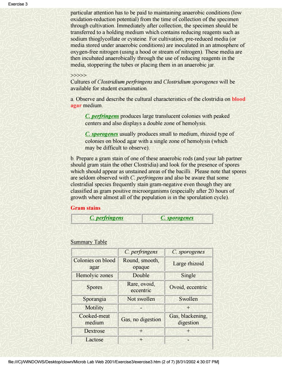
particular attention has to be paid to maintaining anaerobic conditions (low oxidation-reduction potential) from the time of collection of the specimen through cultivation. Immediately after collection, the specimen should be transferred to a holding medium which contains reducing reagents such as sodium thioglycollate or cysteine. For cultivation, pre-reduced media (or media stored under anaerobic conditions) are inoculated in an atmosphere of oxygen-free nitrogen (using a hood or stream of nitrogen). These media are then incubated anaerobically through the use of reducing reagents in the media, stoppering the tubes or placing them in an anaerobic jar. >>>>> Cultures of Clostridium perfringens and Clostridium sporogenes will be available for student examination. a. Observe and describe the cultural characteristics of the clostridia on blood agar medium. C. perfringens produces large translucent colonies with peaked centers and also displays a double zone of hemolysis. C. sporogenes usually produces small to medium, rhizoid type of colonies on blood agar with a single zone of hemolysis (which may be difficult to observe). b. Prepare a gram stain of one of these anaerobic rods (and your lab partner should gram stain the other Clostridia) and look for the presence of spores which should appear as unstained areas of the bacilli. Please note that spores are seldom observed with C. perfringens and also be aware that some clostridial species frequently stain gram-negative even though they are classified as gram positive microorganisms (especially after 20 hours of growth where almost all of the population is in the sporulation cycle). Gram stains C. perfringens C. sporogenes Summary Table C. perfringens C. sporogenes Colonies on blood agar Round, smooth, opaque Large rhizoid Hemolyic zones Double Single Spores Rare, ovoid, eccentric Ovoid, eccentric Sporangia Not swollen Swollen Motility - + Cooked-meat medium Gas, no digestion Gas, blackening, digestion Dextrose + + Lactose + - Exercise 3 file:///C|/WINDOWS/Desktop/clown/Microb Lab Web 2001/Exercise3/exercise3.htm (2 of 7) [8/31/2002 4:30:07 PM]
Exercise 3 Sucrose Salicin Usually- Indole Nitrate production + Gelatin liquification Litmus Milk Test:This assay has not been performed in the lab and will not be included in the lab quiz Bacterial Endospores Bacillus with endospores Endospores are highly resistant resting forms that are produced within the cell Ofthe s ndos s,only two are of medical i and these are the a ngram-positive rods. st spore-forming bacteria are inhabitants of soil,but bacterial spores are found almost everywhere. Because many of the spore-formers can produce quite potent toxins,special precautions must be taken to destroy all spores.Endospores endow the microorganism with increased resistance to heat,irradiation,chemicals,and drying.Endospore formation is a type of cellular differentiation requiring synthesis of specific RNA.proteins.dipicolinic acid.and other components not found in the vegetative cell Upon ta nditions that allo f water,the spore germi nates and differentiates to the vegetative cell Staining of Endospores:Spores produced by bacteria(Bacillus anthracis Clostridium tetani,Clostridium botulinum,e.g)are extremely resistant to desiccation,heat,and chemicals.Ordinary dyes such as crystal violet, safranin,and methylene blue are unable to stain spores because they cannot penetrate the spore coats.These dyes can only stain the vegetative portion of these grar n positive rods,while the endospo remains colorless.Students may perform gram stain on Bacillus species to observe unstained spores file://CWINDOWS/Desktop/clown/Microb Lab Web 2001/Exercise3/exercise3.htm (3 of7)[8/3/002 4:30:07 PM]
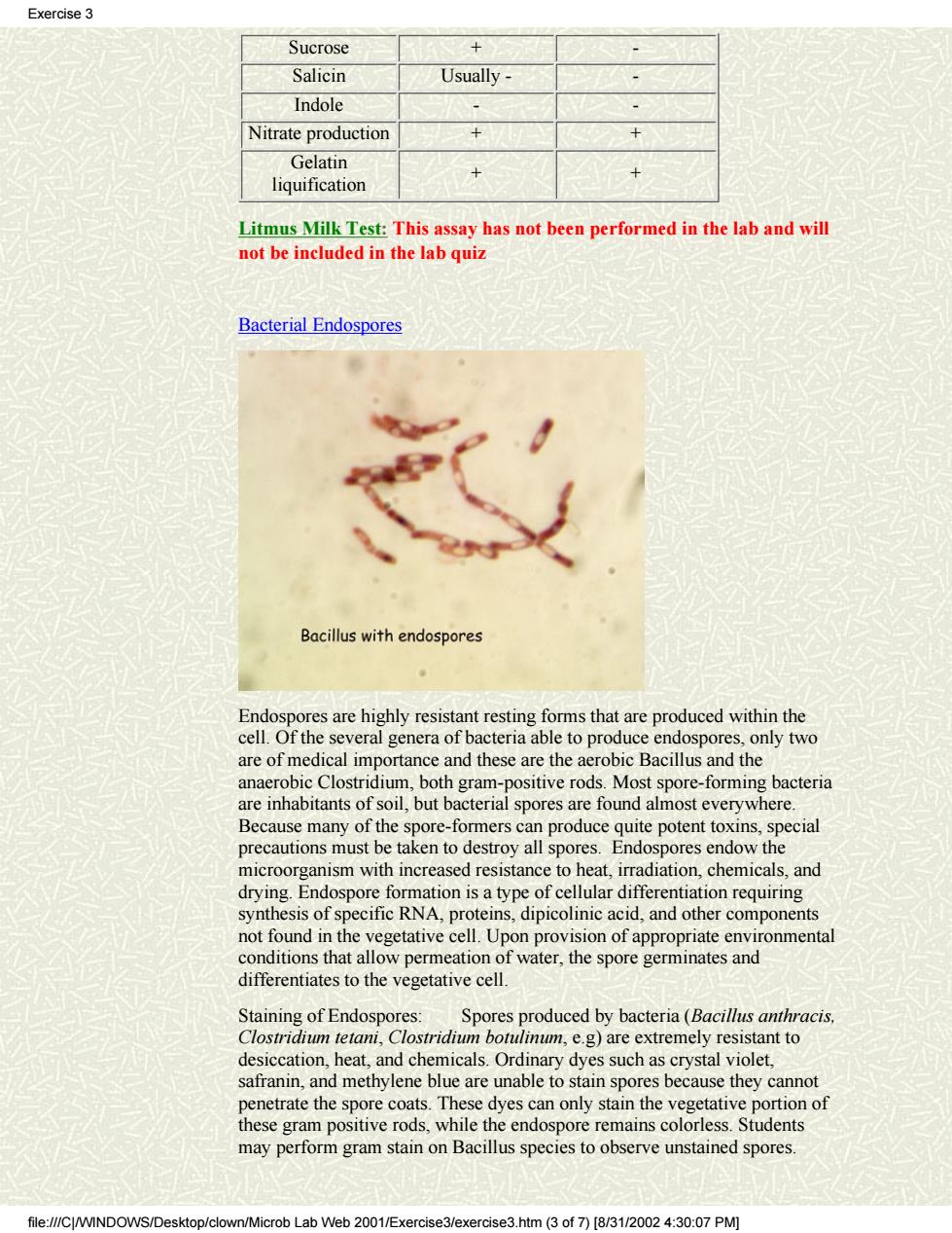
Sucrose + - Salicin Usually - - Indole - - Nitrate production + + Gelatin liquification + + Litmus Milk Test: This assay has not been performed in the lab and will not be included in the lab quiz Bacterial Endospores Endospores are highly resistant resting forms that are produced within the cell. Of the several genera of bacteria able to produce endospores, only two are of medical importance and these are the aerobic Bacillus and the anaerobic Clostridium, both gram-positive rods. Most spore-forming bacteria are inhabitants of soil, but bacterial spores are found almost everywhere. Because many of the spore-formers can produce quite potent toxins, special precautions must be taken to destroy all spores. Endospores endow the microorganism with increased resistance to heat, irradiation, chemicals, and drying. Endospore formation is a type of cellular differentiation requiring synthesis of specific RNA, proteins, dipicolinic acid, and other components not found in the vegetative cell. Upon provision of appropriate environmental conditions that allow permeation of water, the spore germinates and differentiates to the vegetative cell. Staining of Endospores: Spores produced by bacteria (Bacillus anthracis, Clostridium tetani, Clostridium botulinum, e.g) are extremely resistant to desiccation, heat, and chemicals. Ordinary dyes such as crystal violet, safranin, and methylene blue are unable to stain spores because they cannot penetrate the spore coats. These dyes can only stain the vegetative portion of these gram positive rods, while the endospore remains colorless. Students may perform gram stain on Bacillus species to observe unstained spores. Exercise 3 file:///C|/WINDOWS/Desktop/clown/Microb Lab Web 2001/Exercise3/exercise3.htm (3 of 7) [8/31/2002 4:30:07 PM]