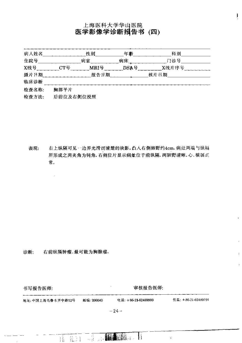
上海医科大学华山医院 医学影像学诊断报告书 (四)》 病人姓名 性别 年转 科别 住院号】 病室 焖宋 门诊号 X线号 CT号 MRI号 DSA号 X线片序号 摄片日期 报告日期 核片日期 格味诊新 检查名称: 州都平片 检查方法: 后前位及右侧位投照 表现: 右上纵隔可见边界光滑而清楚的块影,凸人右侧野约4m病灶两端与纵阔 所形成之两夹角为钝角,右侧位片显示病世位于前纸隔.两肺野清晰,心,横离国 常, 诊断: 名的蚁隔肿蜜,最可能为侧腿棉. 书写报告医师: 审核报告医师: 龙此:中国上海乌静系齐中亮12号 程编:23000 电话:+6216过400 f传森:+621624919到 -24-
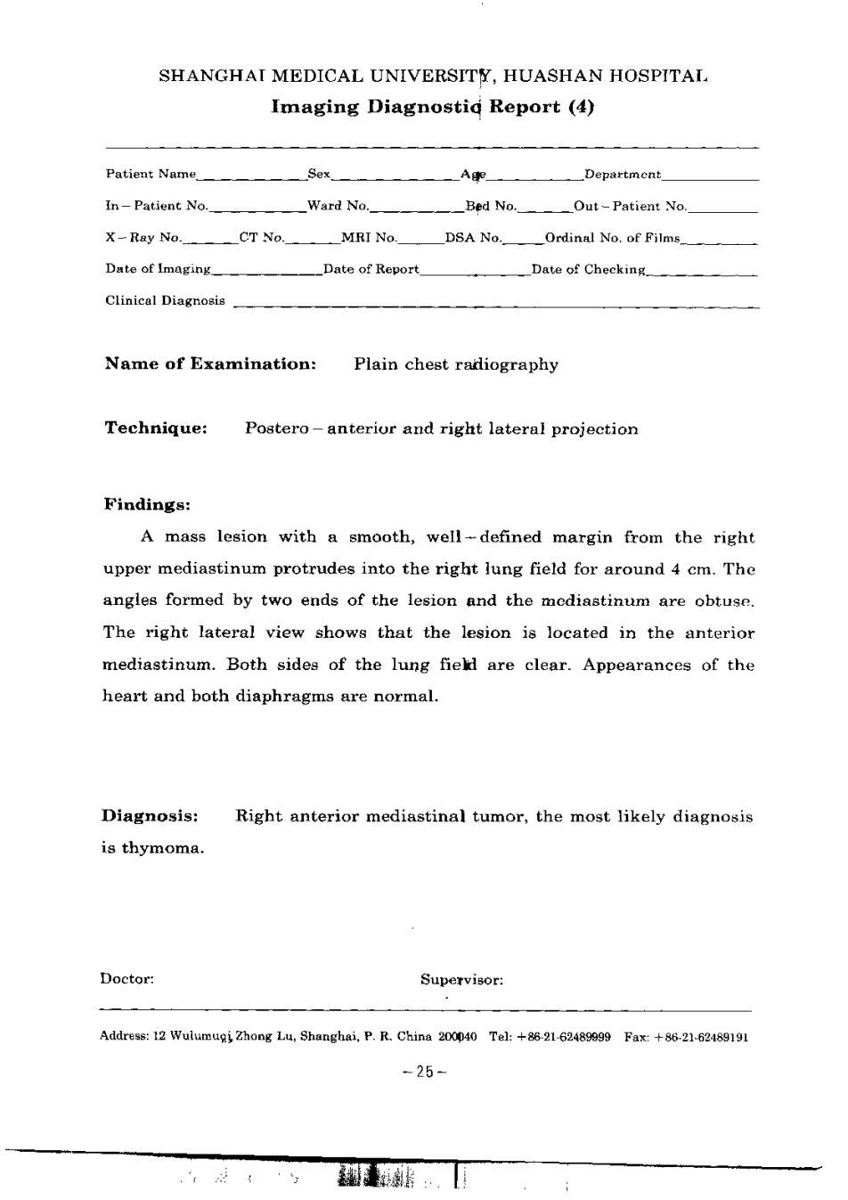
SHANGHAI MEDICAL UNIVERSITY,HUASHAN HOSPITAL Imaging Diagnostid Report (4) Patient Name Sex A Department In-Patient No. Ward No. Bed No. Out-Patient No X-Ray Ne.OT No.MRI No D&A No.Ordinal No.of Pilms Date of Imagang Date of Report Date of Checking. Clinical Diagnosis Name of Examination: Plain chest radiography Technique: Postero-anteriur and right lateral projection Findings: A mass lesion with a smooth,well-defined margin from the right upper mediastinum protrudes into the right lung field for around 4 cm.The angles formed by two ends of the lesion and the modiastinum are obtuse. The right lateral view shows that the lesion is located in the anterior mediastinum.Both sides of the lung field are clear.Appearances of the heart and both diaphragms are normal. Diagnosis对 Right anterior mediastinal tumor,the most likely diagnosis is thymoma. Doctor: Supervisor: Addresse 12 Wulumugi Zhong Lu,Shanghai.P.R.Chins 200090 Tel:+8621-62489099 Fax +86-21-62489191 -25
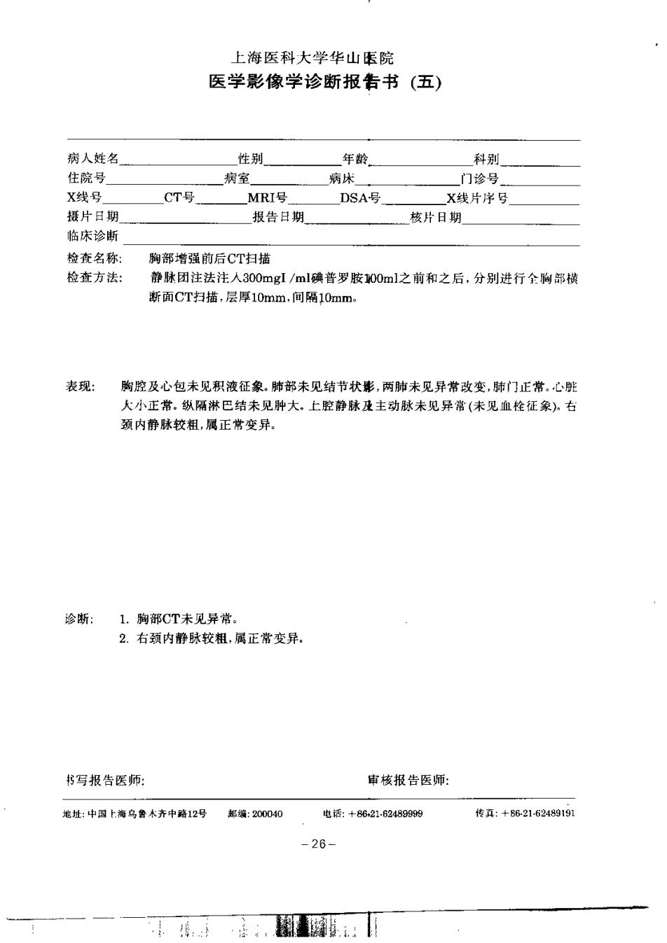
上海医科大学华山玉院 医学影像学诊断报告书(五) 病人姓名 生别 年静 科别 住院号 病室 病休 门诊号」 X线号 CT号 MRI号 DSA号 X线片岸写 摄片日期 报告日期 核片日期 临床诊断 检查名称: 脚部增强前后CT扫描 检查方法: 静琳团注法注人300mgI/ml确若罗胺100ml之前和之后,分别进行全啊部桃 新面CT扫插,层厚10mm.间隔10mm 表现: 胸腔及心包未见积液征象。肺部来见结节状影,两肺未见异常改变,助门正常,心脏 大小正常,纵隔淋巴结未见肿大,上腔静脉是主动脉未见异常(未见血栓任象.右 须内静林较粗,属正鸷变异, 诊断:1.脚部CT未见异常 2.右颈内静脉校组,属正常变异, 书写报告医师: 审核投告医师: 地址:中习上海乌暑木齐中降12号起编:208040 电话:+6621624899附 情真:+8-白148911 -26-
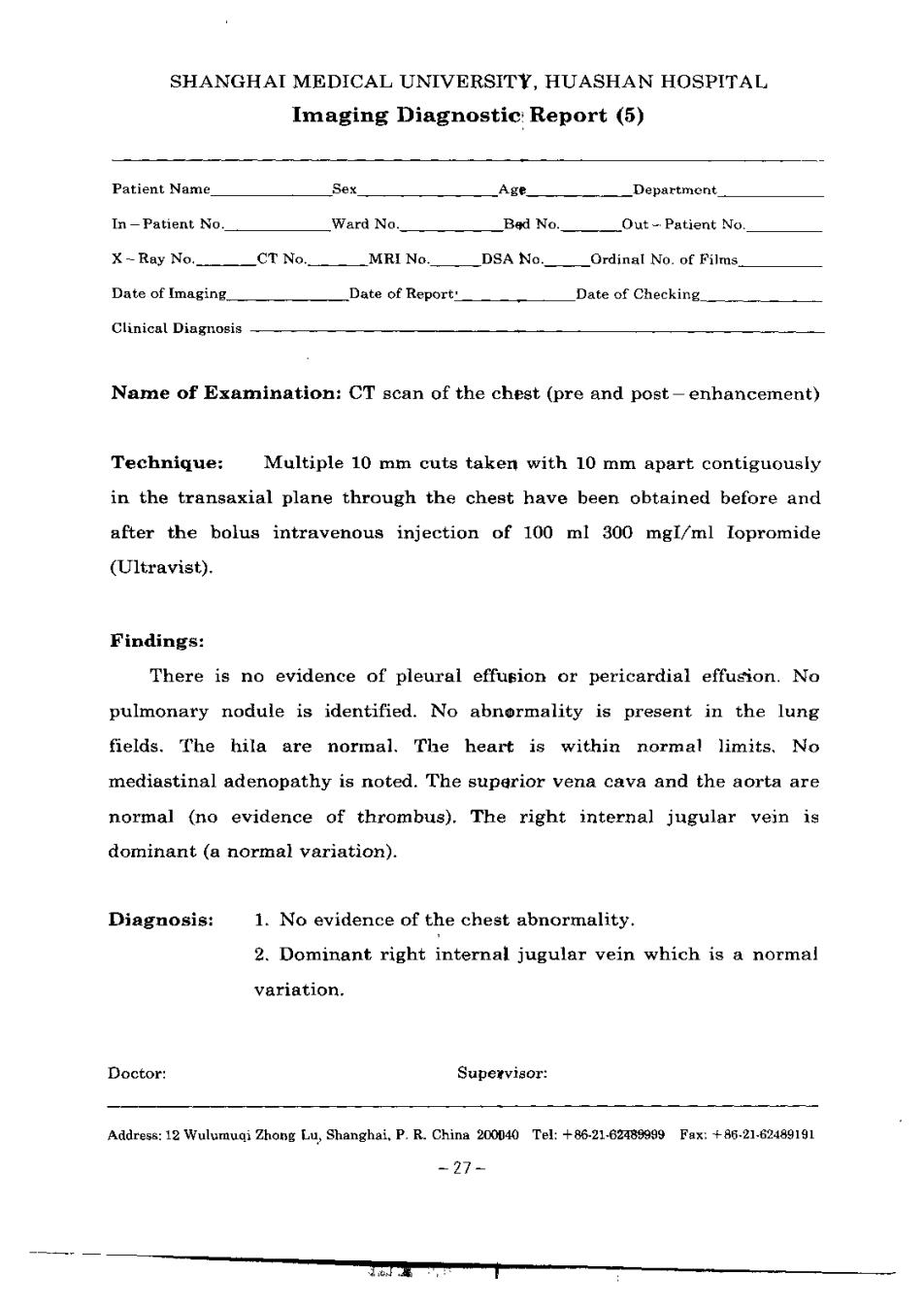
SHANGHAI MEDICAL UNIVERSITY.HUASHAN HOSPITAL Imaging Diagnostic Report (5) Patient Name AcE_ Department In-Patient No. Ward No. Bed No.__Out-Patient No. X-Ray No.CT No..MRI No._DSA No.Ordinal No.of Films Date of Imaging Date of Report: Date of Checking Clinical Dingnosis-—— Name of Examination:CT scan of the chest (pre and post-enhancement) Technique:Multiple 10 mm cuts taken with 10 mm apart contiguously in the transaxial plane through the chest have been obtained before and after the bolus intravenous injection of 100 ml 300 mgl/ml lopromide (Ultraviat). Findings: There is no evidence of pleural effusion or pericardial effusion.No pulmonary nodule is identified.No abnormality is present in the lung fields.The hila are normal.The heart is within normal limits.No mediastinal adenopathy is noted.The superior vena cava and the aorta are normal (no evidence of thrombus).The right internal jugular vein is dominant (a normal variation). Diagnosis: 1.No evidence of the chest abnormality. 2.Dominant right internal jugular vein which is a normal variation. Doetor: Superviaor: Address:12 Wulumuqi Zhong Lo,Shanghai.P.R.China 200040 Tel:+862102 Fax:+86.21-62489191 -27-
上海医科大学华山医院 医学影像学诊断排告书(六) 粥人姓名 性别 年龄 科别 住院号 病室 病床 门诊号」 X线号 CT号 MRI号 D8号」 X线片序号 摄片日期 报告日期 核片日嗣 格床诊新 检在名称: 胸部增强前后CT扫描 检查方法: 采用GE9800CT扫描机作胸部扫猫.静脉团注法注入100m】300mgl/m1拳离 子型造影剂之前和之后,从两肺尖至南侧横辆作陶部连续棍斯断南扫增.丛3 m,问隔3mm.选用的夜.软组织窗和骨前观察. 表现: 右肺底至右上肺野的每一层面上,都可见新月思均匀的水样帝度影与右树壁相 连.此水样密度影的最宽径达4m,右肺受玉,纵隔轻度左移.左肺清嘶。纵隔内本 见异算密度影。 诊新:右测胸腔积液,原因待查。 书写报告医师: 审核报告医师: 始址:中购北海修鲁术齐中群12号车:20040 电话:十8681684680例 传底:+86218216神191 -28-