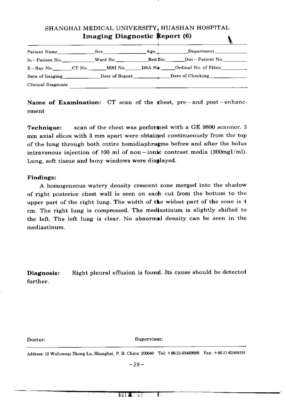
SHANGHAI MEDICAL UNIVERSITY HUASHAN HOSPITAL Imaging Diagnostic Report (6) 麦 Pauent Name Sex Age _DepM代ment In-Patient No Ward No. Bed No. Out-Patient No. XRay No.CT No.MRI No.DSA NeOrdinal No.of Filas. Date of Imaging Dnte of Report Dato of Checking Clinical Diagnosis Name of Examination:CT scan of the chest,pre-and post-enhanc- ement Technique:scan of the cheat was performed with a GE 9800 scanner.3 mm axial slices with 3 mm apart were obtained continueously from the top of the lung through both entire hemidiaphragms before and after the bolus intravenous injection of 100 ml of hon-ionic contrast media (300mgl/ml). Lung.soft tissue and bony windows were displayed. Findings: A homogeneous watery density crescent zone merged into the shadow of right posterior chest wall is seen on each cut from the bottom to the upper part of the right lung.The width of the widest part of the zone is 4 em.The right lung is compressed.The mediastinum is slightly shifted to the left.The left lung is clear.No abnormal density can be seen in the mediastinum. Diagnosis: Right pleural effusion is found.Its cause should be detected further. Doctor: Supervisor: Address:12 Wulumuqi Zhong Lu,Shanghai,P.R.Chirs 200040 Tel:+86-21-63489090 Fax:+86-21-62489191 -29-
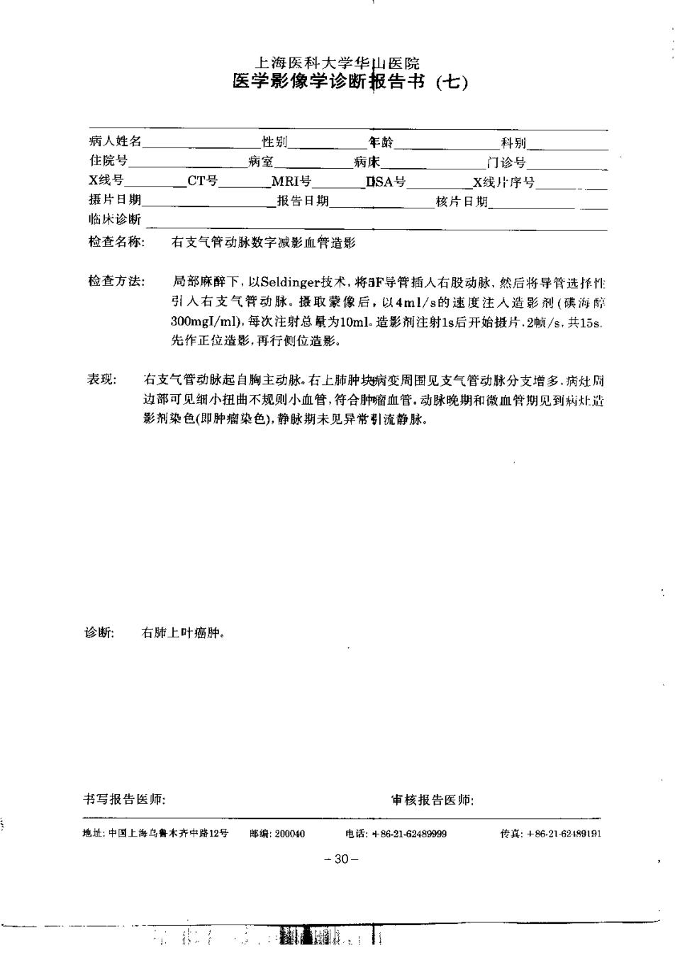
上海医科大学华山医院 医学影像学诊断报告书(七) 病人姓名 性别 午龄 科别 住院号 期室 病床 门诊号 X线号 CT号 MRI号 SA号 X找片序号 摄片目期 报告日期 核片日期 临快珍断 检查名称: 右支气管动称数字减影血件造影 检查方法: 局部麻醉下,以Seldinger技术,将3F学管插人右股动脉,然后将导骨选长性 引入右支气背动肤。摄取蒙像后,以41/s的速度注人是影剂(滨游醇 300mg/ml),每次注射总量为10ml.壶影剂注射1s后开始链片,2就/s,共15s 先作正位造影,再行侧位造影。 表现: 右支气管动脉起白胸主动脉,右上肺肿块谢变周围见支气管动脉分支增多,病灶刷 边部可见细小扭曲不规则小血管,符合肿霜血管,动脉晚期和微血管期见到树灶造 影剂染色(即种榴染色),静肽期未见异君引流静林: 诊断: 右肺上叶密肿, 书写报告医师: 审核报告医师: 然址:中习上海乌鲁木齐中条2号 事编:200040 电话:48际218245999m 传底:◆821621919] -30- ·.:罪.:用
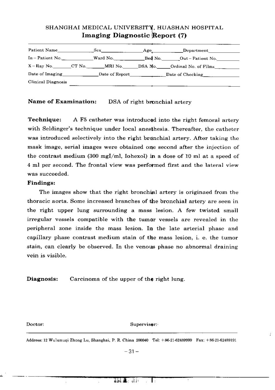
SHANGHAI MEDICAL UNIVERSITY.HUASHAN HOSPITAL Imaging Diagnostic Report (7) Patient Name. Sox_ Age. Department 【n-Patient No. Ward No. Bed No. Out-Patient No. X-Ray No. CT No MRI No.DSA No.Ordinal No.of Films Date of Imaging. Date of Report. Date of Cheeking Clinical Diagnosis Name of Examination: DSA of right bronchial artery Technique:A F5 catheter was introduced into the right femorai artery with Seldinger's technique under local anesthesia.Thereafter,the catheter was introduced selectively into the right branchial artery.After taking the mask image,serial images were obtained one second after the injection of the contrast medium (300 mgI/ml,Iohexol)in a dose of 10 ml at a speed of 4 ml per second.The frontal view was performed first and the lateral view was succeeded. Findings: The images show that the right bronchial artery is originaed from the thoracie aorta.Some increased branches of the bronchial artery are seen in the right upper Iung surrounding a mass lesion.A few twisted small irregular vessels compatible with the tumor veasels are revealed in the peripheral zone inside the mass lesion.In the late arterial phase and capillary phase contrast medium stain of the mass lesion,i.e.the tumor atain,can clearly be observed.In the venous phase no abnormal draining vein is visible. Diagnosis: Careinoma of the upper of the right lung. Doetor: Superviser: Addreas:12 Wulumugi Zhong 1u,Shanghai P.R.China 200040 Tal:+86-2162489999 Fax:+8621-62489191 -31-
读书心得 a而
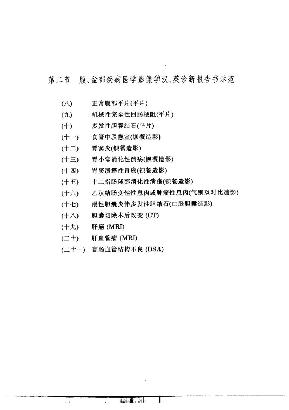
第二节 腹、金部疾病医学影像学汉、英诊新报告书示范 (八) 正常报部平片(平片) (九) 机械性完全性回肠梗阳(平片) (十) 多发性胆囊结石(平片) (十一) 食管中段稳室(钡餐造影) (什三) 骨窦炎(钡餐造影) (什三) 胃小弯消化性遗瑞(钡餐造影) (十四) 肾窦费疡性胃癌(钡餐造影) (十五) 十二指肠球部清化性溃违(钡餐造影) (什六) 乙状结肠变性性息肉或胖瘤性息肉(气钡双对比造影) (十七) 慢性胆囊类伴多发性胆伟石(口服胆囊造影) (什八) 胆囊切除术后改变(CT) (十九)肝癌(MR) (二十) 肝血管瘤MRI) (二十一)盲肠血管结构不良(DSA)