SHANGHAI MEDICAL UNIVERSITY,HUASHAN HOSPITAL Imaging Diagnostic Report (1) Patient Name e% Ae Department In-Patient No Ward No. Bel No._Out-Patient No.. X-Ray No. CT No.MRI No_DSA No_Ordinnl No.of Films_ Date of Imagig Date of Heport Date of Checking Clinieal Diagnosis Name of Examination: Plain chest radiography Technique: Postero-anterior projection Findings: Both sides of the lung field are clear.Shadows of the heart,the diaphragma,the mediastinum,and the visible ribs are nothing remarkable. Diagnosis: Normal P-A chest film. Doctor: Supervisor: Addresec 12 Wulumuqi Zhong Lu,Shanghal,P.R.China 200040 Tel:+86-21-62489090 Fax:+85-21-62489191 -19 理·

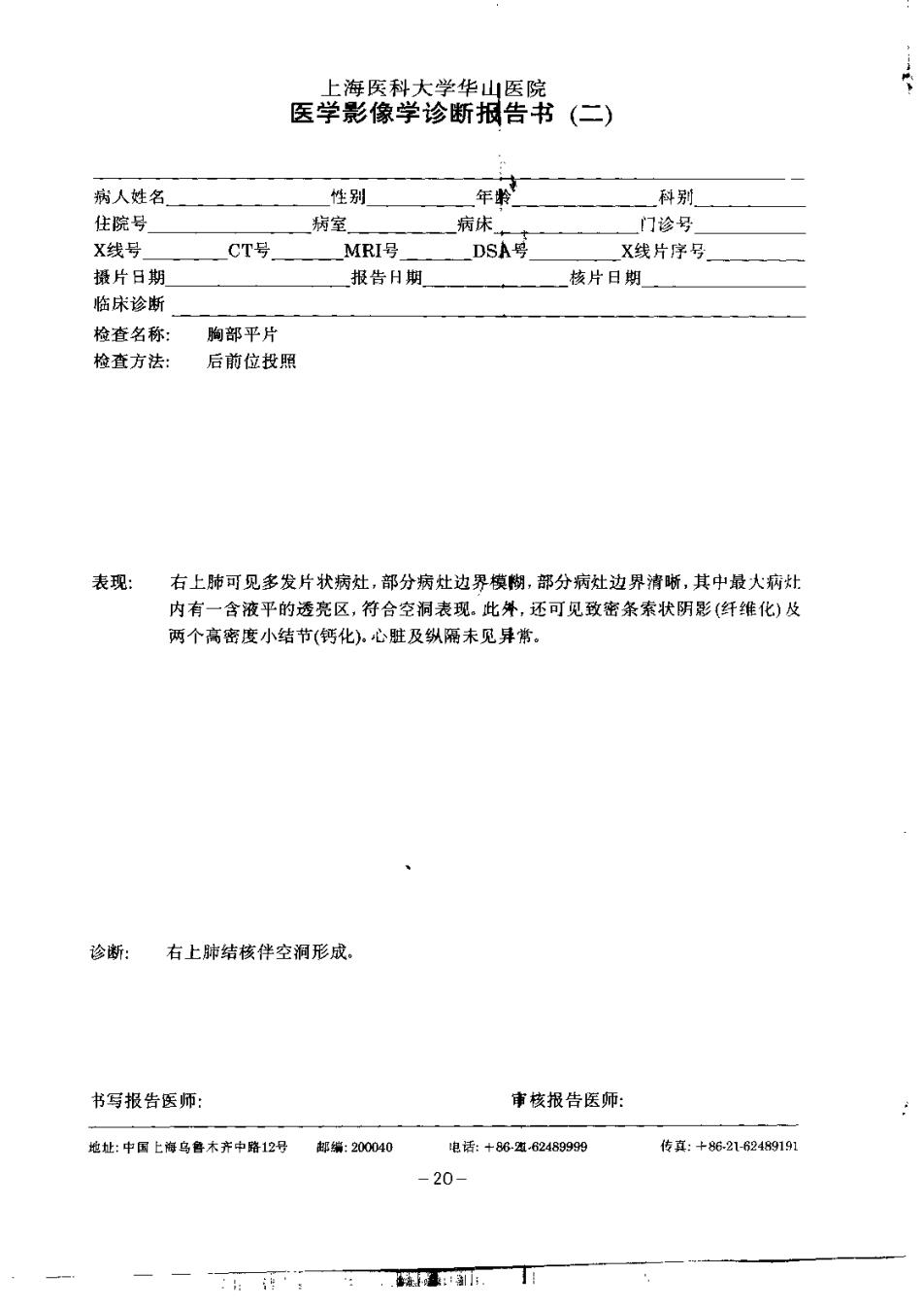
上海医科大学华山医院 医学影像学诊断报告书(二) 病人姓名 性别 年 和别 住院号 病室 病床 门诊号 X线号 CT号 MRI号 DSA号 X线片序号 摄片日期 报告日期 核片日期 临宋诊断 检查名称: 陶部平片 检查方法: 后前位按飘 表现: 右上映可见多发片款病灶,部分病灶边界模胸,部分病灶边界清晰,其中最大病灶 内有一含液平的透亮区,符合空制表现。此弹,还可见致密条素状阴影(纤错化)及 两个高密度小结节(钙化),心脏及纵丽未见异常, 诊断: 右上肺结核伴空洞形成 书写报告医师: 市核报告医师: 途址:中国上海乌鲁木齐中洛12号 都编:200040 电话:+86-看24999阅 转真:+8-216249151 -20-

SHANGHAI MEDICAL UNIVERSITY.HUASHAN HOSPITAL. Imaging Diagnostic Report (2) Patient Nam转 8e× Ag _Dprn专nt In-Patient No. Ward No. Hed No.Out-Patient Nu X一RyNo-—CTNa, MRI No. DSA No.Ordinsl No.of Filme Date of Imaging_ Date of且eport Date of Chocking Clinienl Diagnoais Name of Examination: Plain chest radiography Technique: Postero-anterior projection Findings: Multiple patchy lesions are revealed in the right upper lung.some of them have ill-defined margin and some of them have well-defined margin,inside the largest lesion a round transparent area with a fluid level compatible with a eavity is revealed.Several dark stripes (fibrosis)and two small high density nodules (calcification)are also seen.No abnormality of the heart and the mediastinum is visible. Diagnosis:Pulmonary tuberculosis with cavity formation in the right upper lobe. Doctor: Supervisor: Address:12 Wu'umuqi Zhong Lu.Shanghai,P.R.China 20004 Tek +86-21-63459109 Fax:+862162484191 -21- 上.4·

上海医科大学华中医院 医学影像学诊断报告书(三) 病人姓名 性别 一甲教 科開 住院号 病室 病 门诊号 X线号 CT号 MRI号 D3A号 X线片序号」 摄片日期 报告日期 核片日期 临床诊新 检查名称: 测部平片 检靠方法: 后前位投照及左侧位投赢 表现: 正位侧片见左侧中、上肺野透亮度诚低。与左肺门上方相连.可见-4一5©m大小的 图形块影,其边缘有两个切迹.气件向同侧移位,侧位片上块影之半与静门影重叠, 沿整个前陶竖,即鞠骨后方,可见一5c宽的密度增高带,此高密度带的后,边缘 相当于斜裂,景凹面向后下之蓝线形,右站精帐。 诊断: 左肺门区种块,作左上肺不张,最可能为支气餐肺悠,建议胸部CT检養。 书写报告医师: 审核报告医师: 地址:中偶上特乌鲁本齐中路1空牙 笔编230040 电话:+882182469690 传底:+86216249191 -22-

SHANGHAI MEDICAL UNIVERSITY.HUASHAN HOSPITAL Imaging Diagnostid Report (3) Paticnt Namo Sox In-Patient No. Ward No. Bad No._Out-Patient Na. X-Ray No. CT No.MRI No.DSA No.Ordinal No.of Films Date of Imnging_ Date of Report Dnte of Cheeking Clinical Diagnosis Name of Examination: Plain chest radiography Technique: Postero-anterior and left lateral projection Findings: On the frontal view the transparency of the upper and middle feilds of the left lung is decreased.A 4-5 cm sized round opaque mass lesion with a well-definded margin and two notches is revealed.The medial side of the mass is connected with left upper lung hilum.Ipsilateral (or homolateral) deviation of the trachea is showed.On the lateral film half of the mass lesion overlaps on the shadow of hila.There is a 5 cm wide zone of increased density all the way along the anterior chest wall.behind the sternum.The posterior margin of the high density zone corresponding to the left oblique fiasure is curvilinear with the concaved side faced posteriorly and inferiorly.The right lung field is clear. Diagnosis:A mass lesion connected with the left hilum and loft upper lobe lung collapse is demonstrated.The most likely dingnosis is broncho. genic carcinoma.CT examination of the chest is suggested. Dnetor: Supervisor时 Addres:12 Wulumuqi Zhong Lu.Shanghal,P.R.Chine 200040 Tek +86-21-6249990 Fax:+86-21-82189191 -23- 我