Microscopy:The Instruments 。Refractive index is Unrefracted Oil immersion light objective lens the light-bending ability of a medium. Without immersion oil ·The light may bend in Immersion oil air so much that it misses the small Glass slide high-magnification lens. Condenser lenses 。Immersion oil is used Condenser to keep light from bending. Iris diaphragm Light source Figure 3.3
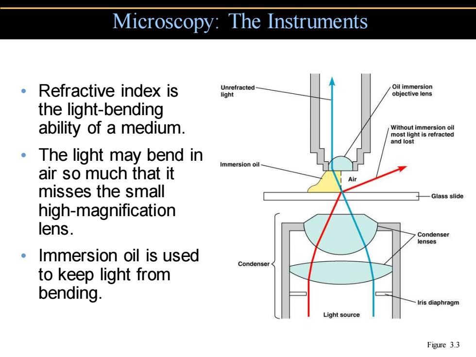
• Refractive index is the light-bending ability of a medium. • The light may bend in air so much that it misses the small high-magnification lens. • Immersion oil is used to keep light from bending. Microscopy: The Instruments Figure 3.3
Brightfield Illumination Eye Eye Ocular lens 。Dark objects are Objective lens Only light reflected by the specimen is visible against a captured by the Specimen objective lens bright background. Unreflected light Condenser lens Light reflected off the Opaque disc specimen does not Light Light enter the objective lens. (a)Brightfield (b)Darkfield Figure 3.4a,b
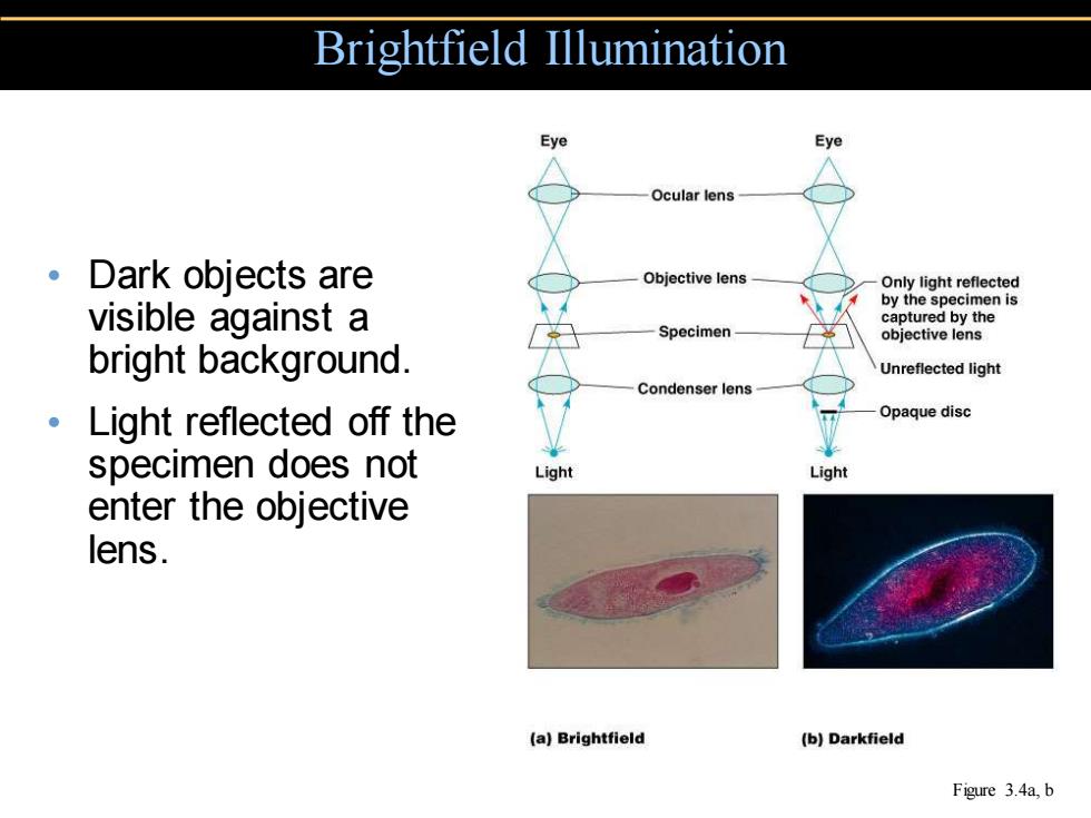
• Dark objects are visible against a bright background. • Light reflected off the specimen does not enter the objective lens. Brightfield Illumination Figure 3.4a, b

Darkfield Illumination Eye Eye Ocular lens Objective lens Only light reflected by the specimen is 。Light objects are captured by the Specimen objective lens visible against a Unreflected light dark background. Condenser lens Opaque disc 。Light reflected off Light Light the specimen enters the objective lens. (a)Brightfield (b)Darkfield Figure 3.4a,b
• Light objects are visible against a dark background. • Light reflected off the specimen enters the objective lens. Darkfield Illumination Figure 3.4a, b

Phase-Contrast Microscopy Eye Ocular lens Diffraction plate Undiffracted light (unaltered by specimen) Objective lens Refracted or diffracted light(altered by specimen) Accentuates diffraction of Specimen Condenser lens the light that passes through a specimen. Annular diaphragm Light (c)Phase-contrast Figure 3.4c
• Accentuates diffraction of the light that passes through a specimen. Phase-Contrast Microscopy Figure 3.4c
Differential Interference Contrast Microscopy 。Accentuates diffraction of the light that passes through a specimen; uses two beams of light. Figure 3.5
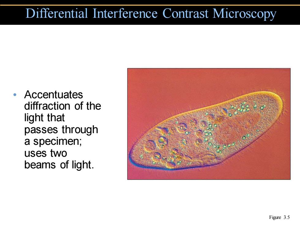
• Accentuates diffraction of the light that passes through a specimen; uses two beams of light. Differential Interference Contrast Microscopy Figure 3.5