(二)荧光显微镜技术 (Fluorescence Microscopy) ?是光镜水平对特异蛋白质等生 -Image plane 物大分子定性定位的有力工具。 Visiple 原理:对样品进行荧光标记, Barrier filter Doc×s transmission of allultraviolet light.allowing 通过激发光激发样品发出荧 -passage of only visible light) 光,只有激发产生的荧光能够 Objective lens 成像,其它光被过滤掉。 Specimen Ultraviolet ?直接荧光标记技术。GFP蛋白 Condenser lens Exciter filter 间接免疫荧光标记技术。 ht and trans Light source Figure A-12 Optics of the Fluorescence Microscope. 主讲:汤华
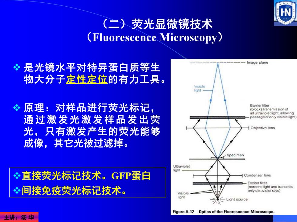
主讲:汤 华 是光镜水平对特异蛋白质等生 物大分子定性定位的有力工具。 原理:对样品进行荧光标记, 通过激发光激发样品发出荧 光,只有激发产生的荧光能够 成像,其它光被过滤掉。 (二)荧光显微镜技术 (Fluorescence Microscopy) 直接荧光标记技术。GFP蛋白 间接免疫荧光标记技术
FLUORESCENT PROBES Dividing cells seen with a fluorescence microscope after staining with specific fluorescent dyes. BH300-FL 荧光显微镜 主讲:汤华
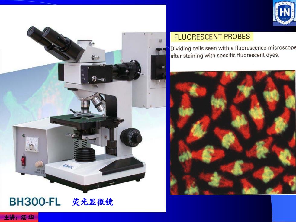
主讲:汤 华 荧光显微镜
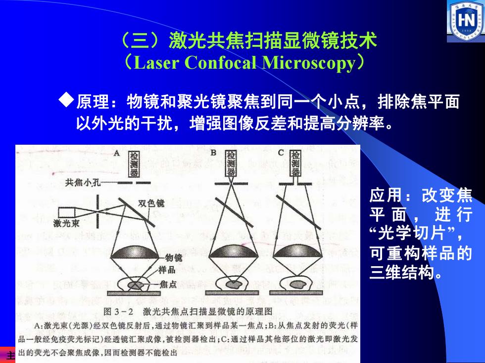
(三)激光共焦扫描显微镜技术 (Laser Confocal Microscopy) ◆原理:物镜和聚光镜聚焦到同一个小点,排除焦平面 以外光的干扰,增强图像反差和提高分辨率。 B 检测器 共焦小孔 双色镜 应用:改变焦 平面,进行 激光束 “光学切片”, 物镜 可重构样品的 样品 三维结构。 达-焦点 图3-2激光共焦点扫描显徽镜的原理图 A:激光束(光源)经双色镜反射后,通过物镜汇聚到样品某一焦点;B:从焦点发射的荧光(样 品一般经免疫荧光标记)经透镜汇聚成像,被检测器检出;C:通过样品其他部位的激光即激光发 出的荧光不会聚焦成像,因而检测器不能检出
主讲:汤 华 (三)激光共焦扫描显微镜技术 (Laser Confocal Microscopy) ¡原理:物镜和聚光镜聚焦到同一个小点,排除焦平面 以外光的干扰,增强图像反差和提高分辨率。 应用:改变焦 平面,进行 “光学切片”, 可重构样品的 三维结构
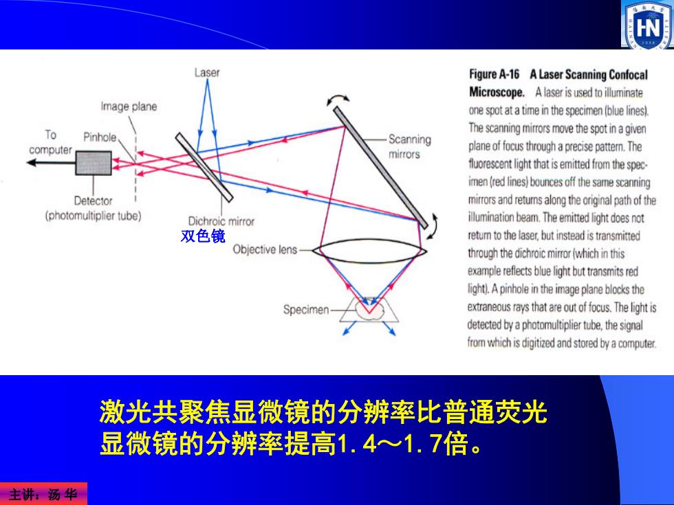
Figure A-16 A Laser Scanning Confocal Microscope.A laser is used to illuminate Image plane one spot at a time in the specimen(blue lines) To The scanning mirrors move the spot in agiven Pinhole Scanning computer mirrors plane of focus through a precise pattem.The fluoresn light that is emitted from the spec- imen (red lines)bounces off the same scanning Detector mirrors and returns along the original path of the (photomultiplier tube) Dichroic mirror illumination beam.The emitted light doesnot 双色镜 retum to the laser,but instead is transmitted Objective lens through the dichroic mirror (which in this example reflects blue light but transmitsred light).A pinhole in the image plane bocks the Specimen extraneous rays that are outof focus.The light is detected by a photomultiplier tube.the signal omwhichisigtidndstored byacmuter. 激光共聚焦显微镜的分辨率比普通荧光 显微镜的分辨率提高1.4心1.7倍。 主讲:汤华
主讲:汤 华 双色镜 激光共聚焦显微镜的分辨率比普通荧光 显微镜的分辨率提高1.4~1.7倍
(四)相差显微镜技术 相差显微镜 (phase contrast microscope):P.Zernike 于1932年发明,并获1953年诺贝尔物理奖。 最大的特点:可以观察未经染色的标本和活细胞。 >基本原理:把透过标本的可见光的光程差变成振幅差,从 而提高了各种结构间的对比度,使各种结构变得清晰可见。 >光线透过标本后发生折射,偏离了原来的光路,同时被延 迟了1/4入(波长),如果再增加或减少1/4入,则光程差变 为1/2入,两束光合轴后干涉加强,振幅增大或减下,提高 反差。 主讲汤华
主讲:汤 华 相差显微镜(phase contrast microscope):由P.Zernike 于1932年发明,并获1953年诺贝尔物理奖。 最大的特点:可以观察未经染色的标本和活细胞。 (四)相差显微镜技术 ¾基本原理:把透过标本的可见光的光程差变成振幅差,从 而提高了各种结构间的对比度,使各种结构变得清晰可见。 ¾光线透过标本后发生折射,偏离了原来的光路,同时被延 迟了1/4λ(波长),如果再增加或减少1/4λ,则光程差变 为1/2λ,两束光合轴后干涉加强,振幅增大或减下,提高 反差