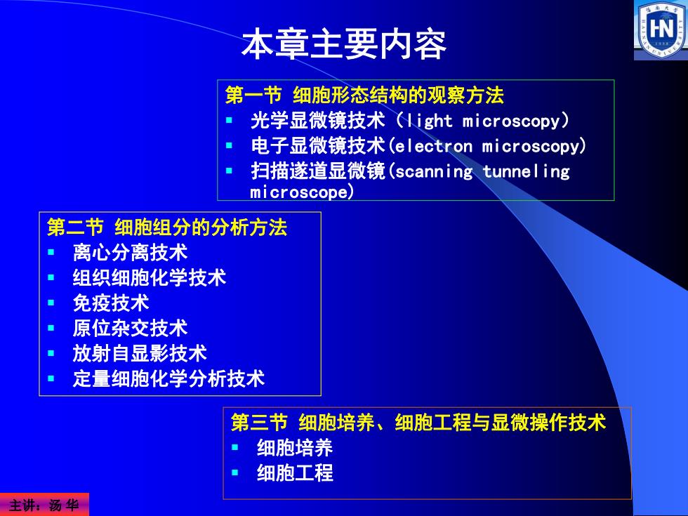
本章主要内容 第一节细胞形态结构的观察方法 光学显微镜技术(light microscopy) 电子显微镜技术(electron microscopy). 扫描遂道显微镜(scanning tunnel ing microscope) 第二节细胞组分的分析方法 离心分离技术 组织细胞化学技术 免疫技术 原位杂交技术 放射自显影技术 定量细胞化学分析技术 第三节细胞培养、细胞工程与显微操作技术 细胞培养 细胞工程 主讲汤华
主讲:汤 华 第一节 细胞形态结构的观察方法 光学显微镜技术(light microscopy) 电子显微镜技术(electron microscopy) 扫描遂道显微镜(scanning tunneling microscope) 第二节 细胞组分的分析方法 离心分离技术 组织细胞化学技术 免疫技术 原位杂交技术 放射自显影技术 定量细胞化学分析技术 第三节 细胞培养、细胞工程与显微操作技术 细胞培养 细胞工程 本章主要内容
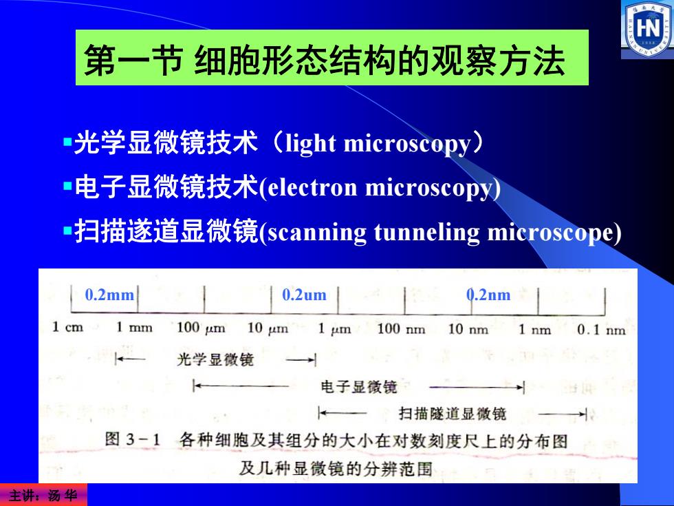
W 第一节细胞形态结构的观察方法 光学显微镜技术(light microscopy) -电子显微镜技术(electron microscopy) -扫描遂道显微镜(scanning tunneling microscope) 0.2mm 0.2um 0.2nm 1cm 1 mm 100m 10m 1 um 100nm 10nm 1 nm 0.1nm 光学显微镜 电子显徽镜 扫描隧道显微镜 图3-1 各种细胞及其组分的大小在对数刻度尺上的分布图 及几种显微镜的分辨范围 主讲汤华
主讲:汤 华 光学显微镜技术(light microscopy) 电子显微镜技术(electron microscopy) 扫描遂道显微镜(scanning tunneling microscope) 第一节 细胞形态结构的观察方法 0.2mm 0.2um 0.2nm
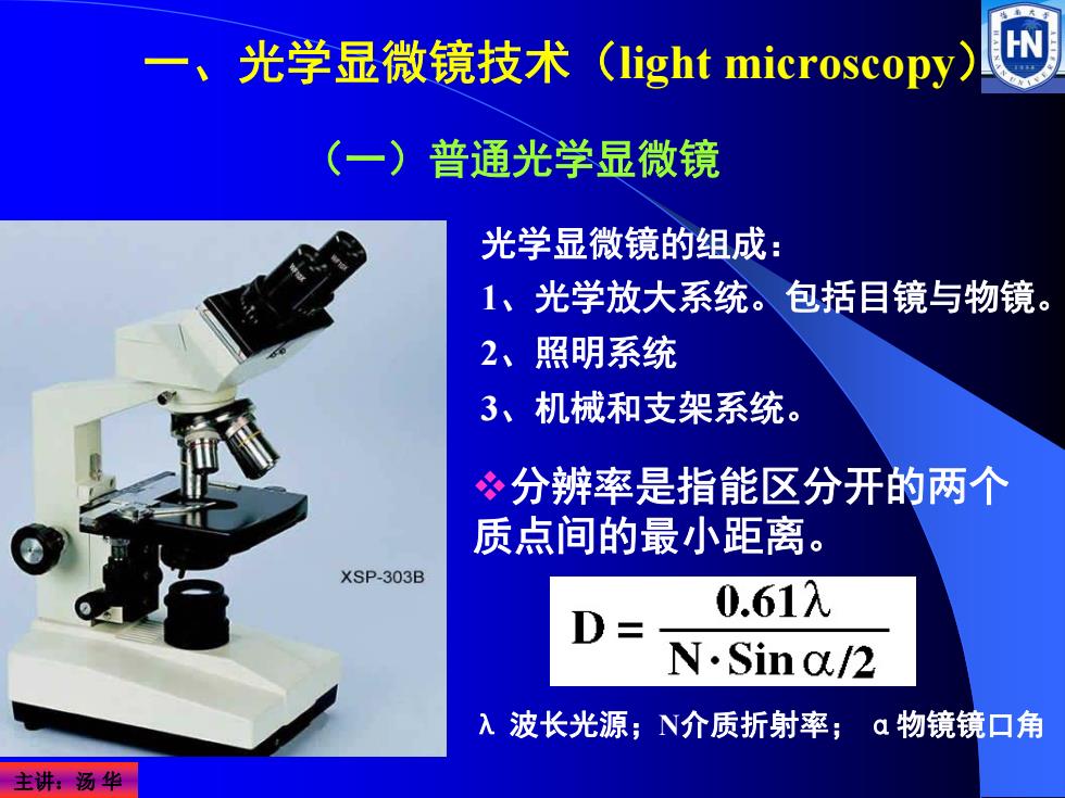
光学显微镜技术(light microscopy) (一)普通光学显微镜 光学显微镜的组成: 1、光学放大系统。包括目镜与物镜。 2、照明系统 3、机械和支架系统。 ?分辨率是指能区分开的两个 质点间的最小距离。 XSP-303B 0.61入 N.Sina/2 入 波长光源;N介质折射率;α物镜镜口角 主讲:汤华
主讲:汤 华 一、光学显微镜技术(light microscopy) 光学显微镜的组成: 1、光学放大系统。包括目镜与物镜。 2、照明系统 3、机械和支架系统。 分辨率是指能区分开的两个 质点间的最小距离。 λ 波长光源;N介质折射率; α物镜镜口角 (一)普通光学显微镜
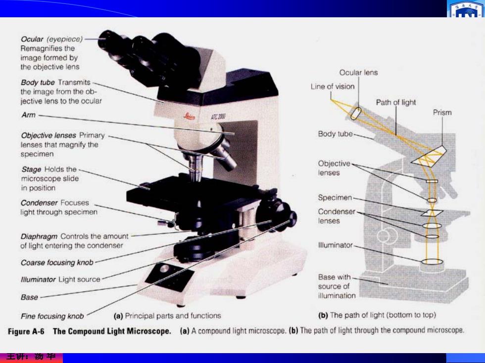
Ocular (eyepiece) Remagnifies the image formed by the objective lens Ocular lens Body tube Transmits Line of vision the image from the ob- jective lens to the ocular Path of light Arm Prism Objective lenses Primary Body tube lenses that magnify the specimen Objective Stage Holds the lenses microscope slide in position Specimen- Condenser Focuses light through specimen Condenser lenses Diaphragm Controls the amount of light entering the condenser Illuminator Coarse focusing knob llluminator Light source- Base with source of Base- illumination Fine focusing knob (a)Principal parts and functions (b)The path of light(bottom to top) Figure A-6 The Compound Light Microscope.(a)A compound light microscope.(b)The path of light through the compound microscope. 王研物甲
主讲:汤 华
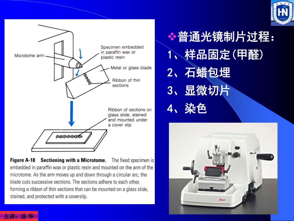
普通光镜制片过程: Specimen embedded in paraffin wax or Microtome arm plastic resin 1、样品固定(甲醛) Metal or glass blade 2、石蜡包埋 Ribbon of thin sections 3、显微切片 Ribbon of sections on 4、染色 glass slide,stained and mounted under a cover slip ccececo Figure A-18 Sectioning with a Microtome.The fixed specimen is embedded in paraffin wax or plastic resin and mounted on the arm of the microtome.As the arm moves up and down through a circular arc,the blade cuts successive sections.The sections adhere to each other, forming a ribbon of thin sections that can be mounted on a glass slide. stained,and protected with a coverslip. 主讲:汤华
主讲:汤 华 普通光镜制片过程: 1、样品固定(甲醛) 2、石蜡包埋 3、显微切片 4、染色