Dental trauma in childhood and adolescence Department of Pediatric Dentistry College of Stomatology, Chongqing Medical University Wang Jin Hua 2010.12.2 Two sections: Section one:general introduction (40 minutes) Section two:dividatur introduction (80 minutes) Contents: Tosp:Clinical classification,diagnositic evidence therapeutic principle and method of dental trauma in childhood and adolescence. To be familiar:Predilection age and type of dental tauma in childhood and Toknow:Incidence of anterior teeth trauma Section one:general introduction 1.1 Definition.1.2 Epidemiology.1.3 Classification.1.4 History and examination. Childhood and adolescence teeth suffered sharp trauma,especially dental,pulp. periodontium trauma due to beating and collisions. 1.2 Epidemiology Table 1 dental trauma prevalence in childhood and adolescence. age Prevalence Boys girls Five vears group 31-40% 16-30% Twelve vears group 12.330% 4-19% Table2 dental trauma incidence in childhood and adole dental trauma incidence Primary dentition Permanent dentition predilection age 1-2 vears 7-9 ears ccount ratio 50% 50-700% Table marison tothe Primary dentition and Permanent dentition clinical comparison to the Primary dentition and Permanent dentition Primary dentition trauma Permanent dentition trauma Displacement is common Tooth fracture is common ntrusion.extrusion,displacement Muturation of fracture or roo fracture Immuturation of root:mobility,displacement,extrusion Account 80%in Primary trauma Account 40-60%in Permanent trauma
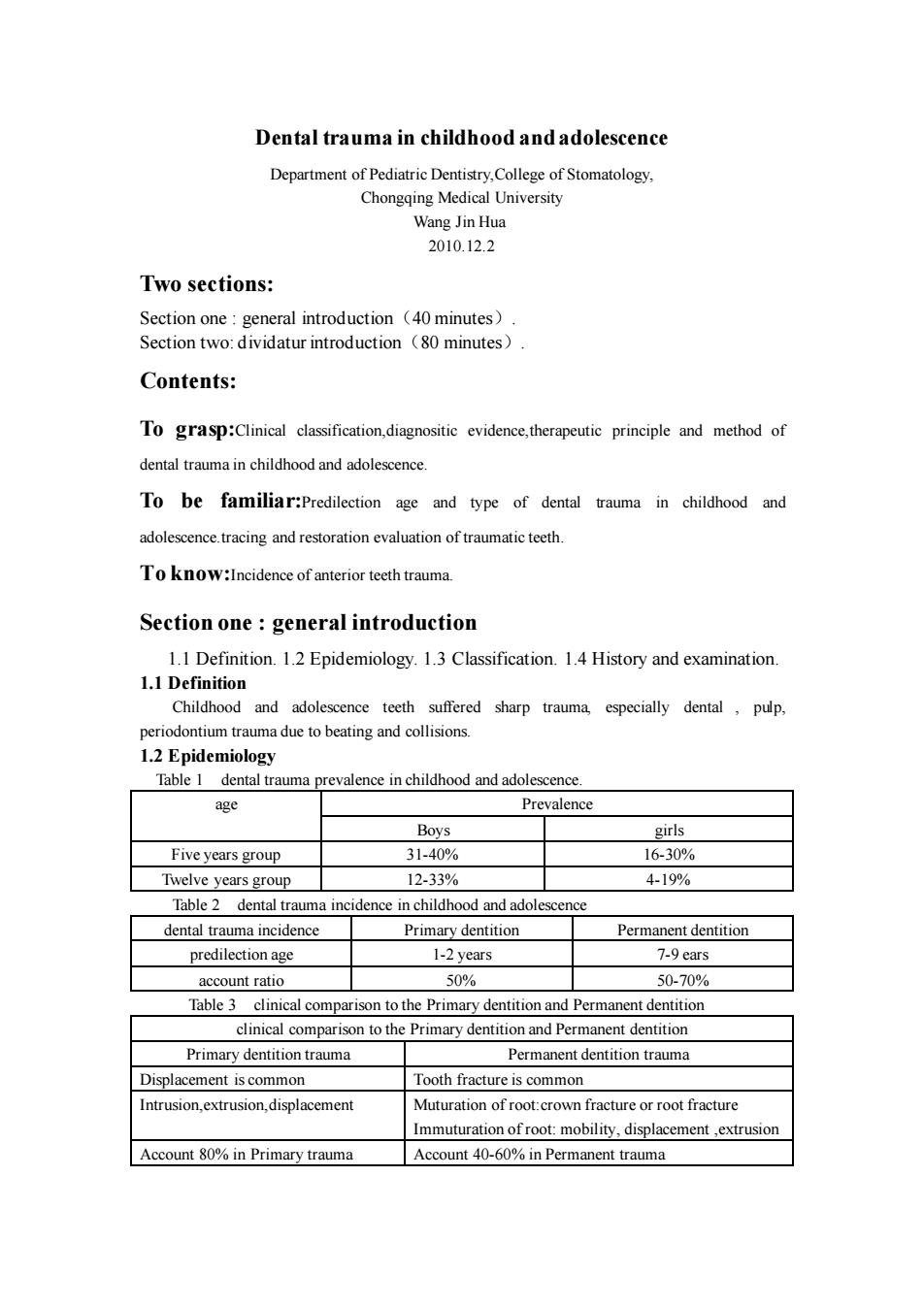
Dental trauma in childhood and adolescence Department of Pediatric Dentistry,College of Stomatology, Chongqing Medical University Wang Jin Hua 2010.12.2 Two sections: Section one : general introduction(40 minutes). Section two: dividatur introduction(80 minutes). Contents: To grasp:Clinical classification,diagnositic evidence,therapeutic principle and method of dental trauma in childhood and adolescence. To be familiar:Predilection age and type of dental trauma in childhood and adolescence.tracing and restoration evaluation of traumatic teeth. To know:Incidence of anterior teeth trauma. Section one : general introduction 1.1 Definition. 1.2 Epidemiology. 1.3 Classification. 1.4 History and examination. 1.1 Definition Childhood and adolescence teeth suffered sharp trauma, especially dental , pulp, periodontium trauma due to beating and collisions. 1.2 Epidemiology Table 1 dental trauma prevalence in childhood and adolescence. age Prevalence Boys girls Five years group 31-40% 16-30% Twelve years group 12-33% 4-19% Table 2 dental trauma incidence in childhood and adolescence dental trauma incidence Primary dentition Permanent dentition predilection age 1-2 years 7-9 ears account ratio 50% 50-70% Table 3 clinical comparison to the Primary dentition and Permanent dentition clinical comparison to the Primary dentition and Permanent dentition Primary dentition trauma Permanent dentition trauma Displacement is common Tooth fracture is common Intrusion,extrusion,displacement Muturation of root:crown fracture or root fracture Immuturation of root: mobility, displacement ,extrusion Account 80% in Primary trauma Account 40-60% in Permanent trauma
13 Classification (f-study) Andreasen classification(self-study) Li hong yi classification (focal point) Table 4 classification of the dental trauma in childhood and adolescence(Li hong yi classification) Periodontal injury Pulpal injury Dental iniury Crown fracture Tooth fracture Root fracture Crown-root fracture intrusion Displacement Lateral luxation Part luxation Total extrusion 1.4 History and examination 1.4.1 Dental history 1.When did injury occur? 2.Where did injury occur? 3.How did iniury occur? 4.Concussion,headache,vomiting or amnesia? 1.4.2 Clinical examination Facial swelling.bruises,mandibular movement or mandibular Inspection deviation on opening or closing the mouth crowr fracture.mobility Palpation and percussion sensitive to percussion,displacement,cinch,roo fracture,alveola fracrute,and so on Pulpal examination Thermal tests,cold tests,electric puip tests,neither negative nor (reaction to sensitivity tests) positive responses should be trusted immediately after trauma. 1.4.3 Radiographic examination I.Crown and root fracture,and their condition. 2.Alveolar and jaw bones fracrute,and their condition. 3.Condition of the periodontal tissue. 4.Condition of adja tand dental germ Condionofrotformaionandabsorption 6. Relationship between primary teeth and succeed permanent teeth 7.If there are root absorption and modus to the old trauma
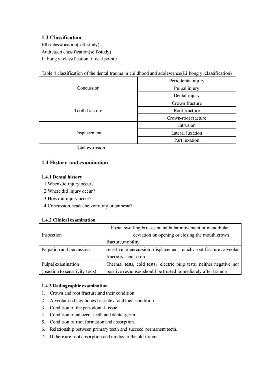
1.3 Classification Ellis classification(self-study). Andreasen classification(self-study) Li hong yi classification(focal point) Table 4 classification of the dental trauma in childhood and adolescence(Li hong yi classification) Concussion Periodontal injury Pulpal injury Dental injury Tooth fracture Crown fracture Root fracture Crown-root fracture Displacement intrusion Lateral luxation Part luxation Total extrusion 1.4 History and examination 1.4.1 Dental history 1.When did injury occur? 2.Where did injury occur? 3.How did injury occur? 4.Concussion,headache,vomiting or amnesia? 1.4.2 Clinical examination Inspection Facial swelling,bruises,mandibular movement or mandibular deviation on opening or closing the mouth,crown fracture,mobility Palpation and percussion sensitive to percussion,displacement,cinch,root fracture,alveolar fracrute,and so on. Pulpal examination (reaction to sensitivity tests) Thermal tests, cold tests,electric puip tests, neither negative nor positive responses should be trusted immediately after trauma. 1.4.3 Radiographic examination 1. Crown and root fracture,and their condition. 2. Alveolar and jaw bones fracrute,and their condition. 3. Condition of the periodontal tissue. 4. Condition of adjacent teeth and dental germ. 5. Condition of root formation and absorption. 6. Relationship between primary teeth and succeed permanent teeth. 7. If there are root absorption and modus to the old trauma
Section two:dividatur introduction 2.1 concussion.2.2 tooth fracture. 2.3 displacement First paragraph:Concussion 1.Definitionto effect periodontal tissue and pulp tissue chiefy after tooth trauma.but tooth sue Integrity or display to be enamel thereare o serous tissues defect and tooth ation 2.Clinical manifestation: Periodontal tissue injury pulp tissue injury:dental tissue injury 2.1 Periodontal tissue injury Tooth aching Percussion unwell or sensitive to percussion. Radiograph:no abnormalities or periodontal spaces widen. 2.2 Pulp tissue injury estion bleed Ser change of pulp traumatic kystis,root paraplasia 2.3 Dental tissue injury Enamel infraction 3.Treatment Eliminat occlusion trauma (modulate occlusion) Diminish or avoid irritation (refrain from caloric stimulation and biting) Infection prevention(take anti-inflammatory) Deal with enamel surface infraction (apply fluorine varnish) Follow-up review periodic(may be one week then one month and so on) second paragraph:Tooth fracture 1.Definition:tooth sclerous tissues fracture,take place in enamel,dentin.cementum 22 Root fracture 2 3 Crown-root fracture 2.1 Crown fracture:it was the commonest in the othfracture,it was predilected inincisa ang and incisal edge It was divided into merely enamel fracture,enamel fracture with dentin exposed crown fracture with pulp exposed 2.1.1 Merely enamel fracture Clinic:twas predilected in incisa angle and incisa edgeand dentin didn't expos Treatment: No disposal. Grinding and polishing To restore in the near future for the small areas
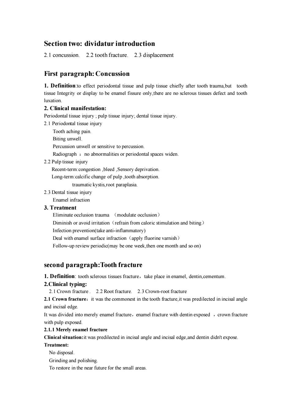
Section two: dividatur introduction 2.1 concussion. 2.2 tooth fracture. 2.3 displacement First paragraph:Concussion 1. Definition:to effect periodontal tissue and pulp tissue chiefly after tooth trauma,but tooth tissue Integrity or display to be enamel fissure only,there are no sclerous tissues defect and tooth luxation. 2. Clinical manifestation: Periodontal tissue injury ; pulp tissue injury; dental tissue injury. 2.1 Periodontal tissue injury Tooth aching pain. Biting unwell. Percussion unwell or sensitive to percussion. Radiograph :no abnormalities or periodontal spaces widen. 2.2 Pulp tissue injury Recent-term:congestion ,bleed ,Sensory deprivation. Long-term:calcific change of pulp ,tooth absorption. traumatic kystis,root paraplasia. 2.3 Dental tissue injury Enamel infraction 3. Treatment Eliminate occlusion trauma (modulate occlusion) Diminish or avoid irritation(refrain from caloric stimulation and biting) Infection prevention(take anti-inflammatory) Deal with enamel surface infraction(apply fluorine varnish) Follow-up review periodic(may be one week,then one month and so on) second paragraph:Tooth fracture 1. Definition: tooth sclerous tissues fracture,take place in enamel, dentin,cementum. 2.Clinical typing: 2.1 Crown fracture . 2.2 Root fracture. 2.3 Crown-root fracture 2.1 Crown fracture:it was the commonest in the tooth fracture,it was predilected in incisal angle and incisal edge. It was divided into merely enamel fracture,enamel fracture with dentin exposed ,crown fracture with pulp exposed. 2.1.1 Merely enamel fracture Clinical situation:it was predilected in incisal angle and incisal edge,and dentin didn't expose. Treatment: No disposal. Grinding and polishing. To restore in the near future for the small areas
To restore after viewing for the large areas essential to pulp protection. Treatment: Took indireet pulp capping to protect pulp Mobility not obviously:direct repair mobility vanishing Modulate occlusion Follow-up review periodic 2.1.3 Crown fracture with pulp exposed Clinical situ hpulp pod,theyeresntive to the caloric d.it could be infected and pulp hyperblastosis if it didn't treat. Treatment: Take direct pulp capping(it was difficult to success). Take palpotomy(root immaturating). Take apexifica Take root canal therapy(o maturating) 2.2 Root fracture:It occur most frequently in the middle or the apical third of the root we can diagnose by radiograph and varying projection angle. 2.2.1 It can be divided into three types: fracture. apical third root fracture Tooth mobility;dental crown lengthen slightly;articulation trauma 2.2.3 Therapeutic principle Reset to broken ends of fractured root.:splint tooth(2-3mouths):eliminate occlusion trauma. 2 2.4 Therany method Coronal third: xtract the fragment and retain the remaining root by extirpate gingiv and strip alveolar bone partly.if the root was immture.we can Take apexification.keep the teeth interspace ,and Take root canal therapy when the root was mature,if the root was mature,we take root canal therapy directly,then draw root out of alveolar crest.this is root canal-orthodontic treatment clinical prognosis was worse Middle third: ent fixed for-3mouths then heaing and pulp vitality reovery.iftake place therapy when broken ends healing,otherwise we place retention post in root canal Apical third:prognosis is better than other root fracture,if the root fracture is almost immobility.we can let alone if there are mobility obviously we can fix it:if take place root tip 2.3 Crown-root f usly when teeth traum take place,it can be divided into two types:simple crown-root fracture and complicated crown-roo fracture. 2.3.1 Clinical manifestation Horizontal fracture and vertical fracture
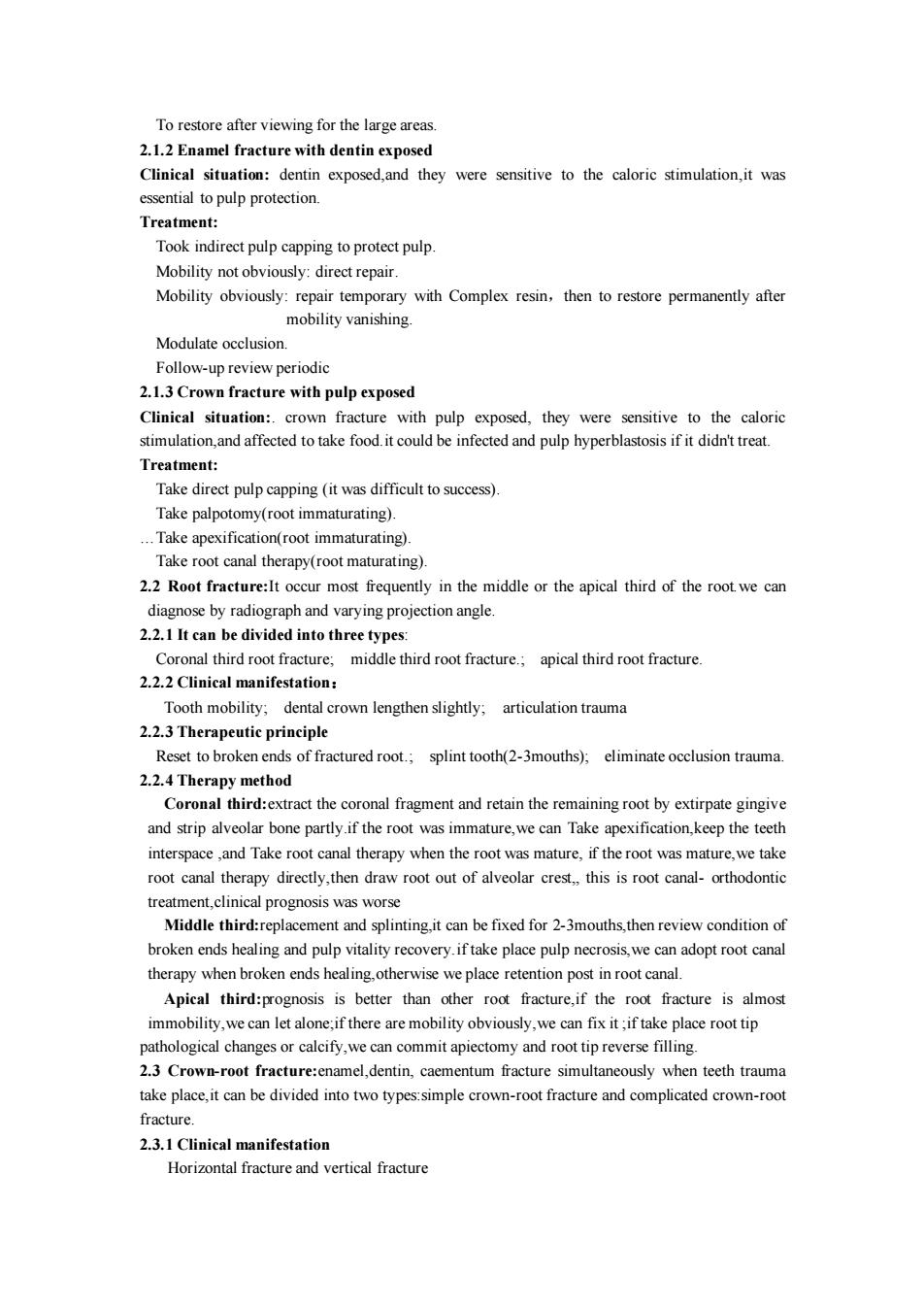
To restore after viewing for the large areas. 2.1.2 Enamel fracture with dentin exposed Clinical situation: dentin exposed,and they were sensitive to the caloric stimulation,it was essential to pulp protection. Treatment: Took indirect pulp capping to protect pulp. Mobility not obviously: direct repair. Mobility obviously: repair temporary with Complex resin,then to restore permanently after mobility vanishing. Modulate occlusion. Follow-up review periodic 2.1.3 Crown fracture with pulp exposed Clinical situation:. crown fracture with pulp exposed, they were sensitive to the caloric stimulation,and affected to take food.it could be infected and pulp hyperblastosis if it didn't treat. Treatment: Take direct pulp capping (it was difficult to success). Take palpotomy(root immaturating). .Take apexification(root immaturating). Take root canal therapy(root maturating). 2.2 Root fracture:It occur most frequently in the middle or the apical third of the root.we can diagnose by radiograph and varying projection angle. 2.2.1 It can be divided into three types: Coronal third root fracture; middle third root fracture.; apical third root fracture. 2.2.2 Clinical manifestation: Tooth mobility; dental crown lengthen slightly; articulation trauma 2.2.3 Therapeutic principle Reset to broken ends of fractured root.; splint tooth(2-3mouths); eliminate occlusion trauma. 2.2.4 Therapy method Coronal third:extract the coronal fragment and retain the remaining root by extirpate gingive and strip alveolar bone partly.if the root was immature,we can Take apexification,keep the teeth interspace ,and Take root canal therapy when the root was mature, if the root was mature,we take root canal therapy directly,then draw root out of alveolar crest, this is root canal- orthodontic treatment,clinical prognosis was worse Middle third:replacement and splinting,it can be fixed for 2-3mouths,then review condition of broken ends healing and pulp vitality recovery.if take place pulp necrosis,we can adopt root canal therapy when broken ends healing,otherwise we place retention post in root canal. Apical third:prognosis is better than other root fracture,if the root fracture is almost immobility,we can let alone;if there are mobility obviously,we can fix it ;if take place root tip pathological changes or calcify,we can commit apiectomy and root tip reverse filling. 2.3 Crown-root fracture:enamel,dentin, caementum fracture simultaneously when teeth trauma take place,it can be divided into two types:simple crown-root fracture and complicated crown-root fracture. 2.3.1 Clinical manifestation Horizontal fracture and vertical fracture
Horizontal fracture:it is common in clinical,display med-distal direction,dental crown labial ace droop partly lingual o with gingive sometimes my be takeo gingive and pulp stimulusaching Vertical fracture:there isonly single band fracture line usually,and it is few in clinical 2.3.2 Therapy method Emergency treatment:fixed temporary Simple crown-roo fracture:cemn repair stickiness treatment Systemical therapy: Reparation after removing dental crown fragment. Remove dental crown fragment and assist gingivectomy and remove alveolar bone. Root canal-orthodontic combined treatment Third paragraph:Tooth displacement 1.Definision:when outside force contributed teeth,it made the teeth separate from normal nosition be divided into three type 2.2part extrusion:2.3total extrusion 2.1 Tooth intrusion 2.1.1 Definision:when outside force contributed teeth,the teeth were inserted alveolar bone 2.1.2 Clinical manifestation It was shorter than the adiacent teeth and no mobility the inrusive teeth and the just eruptive teeth. Percussion:intrusive teeth take on low and deep tone.but the just eruptive teeth present high metal tone. Radiograph:the intrusive teeth take on image vanishing of periodontal spaces and laminadura.but the just eruptive teeth precent nommal image of periodontal spaces and lamina dura 2.1.3 Treatment Root tip immaturation:re-erupted spontaneously.but when the apical shadow and the apical root resorption happened,we should extirpate pulp.then apply calcium hydroxide to obturatie the root canal Root tip maturation:orthodontic hook,and take root canal therapy after teeth trauma. Only root tip immature teeth can be vital pulp Root resorption:root tip immature teeth:58%:root tip mature teeth:70% 2.2 Lateral luxation and part extrusion 22 1 Definision. Lateral luxation:when outside force contributed the teeth,the teeth take on ligula Part extrusion:when outside force contributed teeth,the teeth present to be prolapsed out of tooth socket and lengthen obviously 2.2.2 Clinical manifestation
Horizontal fracture:it is common in clinical,display med-distal direction,dental crown labial surface droop partly,lingual surface connected with gingive sometimes.it may be take on gingive and pulp stimulus aching. Vertical fracture:there is only single band fracture line usually,and it is few in clinical. 2.3.2 Therapy method Emergency treatment:fixed temporary. Simple crown-root fracture: cemental repair. Vertical fracture: stickiness treatment. Systemical therapy: Reparation after removing dental crown fragment. Remove dental crown fragment and assist gingivectomy and remove alveolar bone. Root canal- orthodontic combined treatment. Third paragraph:Tooth displacement 1.Definision:when outside force contributed teeth,it made the teeth separate from normal position. 2.It can be divided into three types 2.1intrusion; 2.2lateral luxation; 2.2part extrusion; 2.3total extrusion. 2.1 Tooth intrusion 2.1.1 Definision: when outside force contributed teeth,the teeth were inserted alveolar bone. 2.1.2 Clinical manifestation It was shorter than the adjacent teeth,and no mobility. We pay attention to the difference between the intrusive teeth and the just eruptive teeth. Percussion:intrusive teeth take on low and deep tone,but the just eruptive teeth present high metal tone. Radiograph:the intrusive teeth take on image vanishing of periodontal spaces and laminadura.but the just eruptive teeth precent normal image of periodontal spaces and lamina dura. 2.1.3 Treatment Root tip immaturation:re-erupted spontaneously.but when the apical shadow and the apical root resorption happened ,we should extirpate pulp,then apply calcium hydroxide to obturatie the root canal. Root tip maturation: orthodontic hook,and take root canal therapy after teeth trauma. 2.1.4 Prognosis Only root tip immature teeth can be vital pulp. Root resorption: root tip immature teeth:58%; root tip mature teeth:70%. 2.2 Lateral luxation and part extrusion 2.2.1 Definision: Lateral luxation:when outside force contributed the teeth,the teeth take on ligula dislocation,distoversion and mesioversion. Part extrusion: when outside force contributed teeth,the teeth present to be prolapsed out of tooth socket and lengthen obviously. 2.2.2 Clinical manifestation