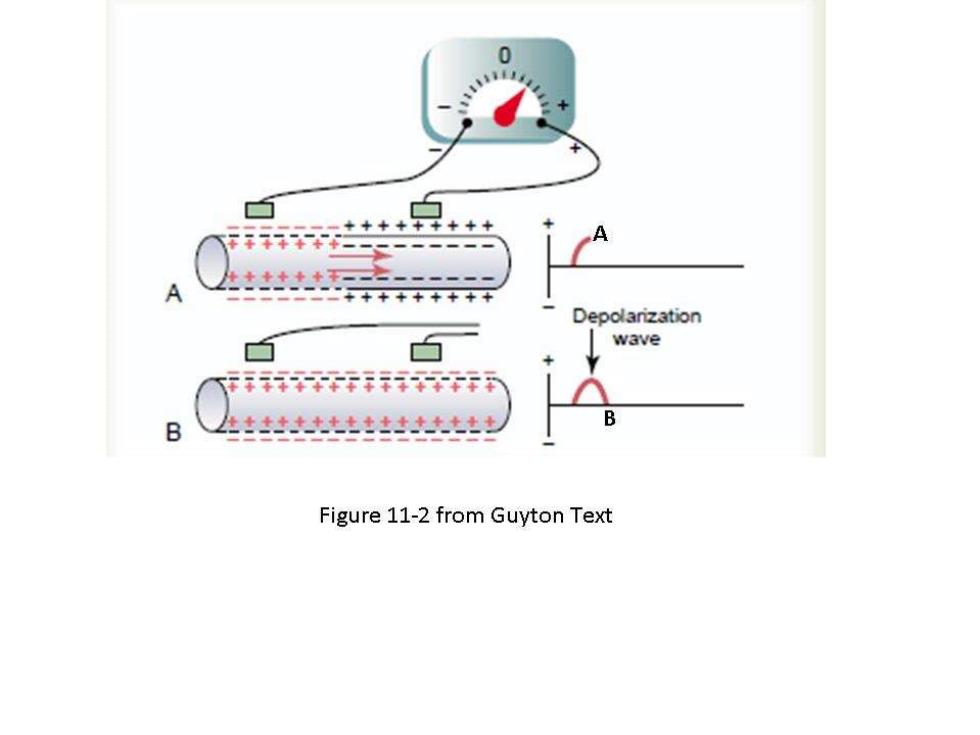
0 +++++++++ Depolarization 白 wave 行行行用 B Figure 11-2 from Guyton Text
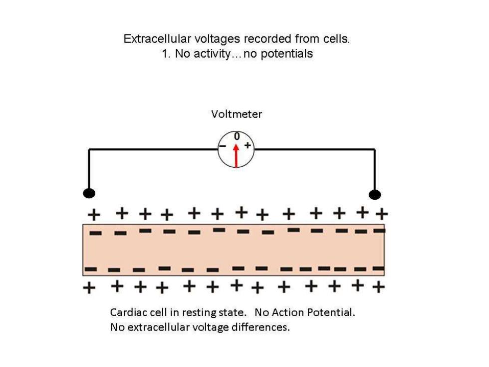
Extracellular voltages recorded from cells. 1.No activity...no potentials Voltmeter ++++++++++++ 十 ■ ■一■■■■■■ +++++++++++++ Cardiac cell in resting state.No Action Potential. No extracellular voltage differences
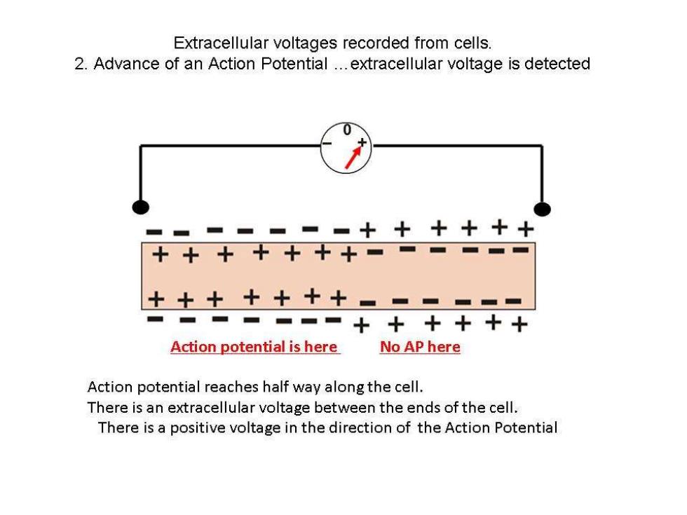
Extracellular voltages recorded from cells. 2.Advance of an Action Potential...extracellular voltage is detected ▣===+十十+十十 十++++十+■一==▣▣ 十十十十十十十====== ■-■■■一■十++十+十 Action potential is here No AP here Action potential reaches half way along the cell. There is an extracellular voltage between the ends of the cell. There is a positive voltage in the direction of the Action Potential
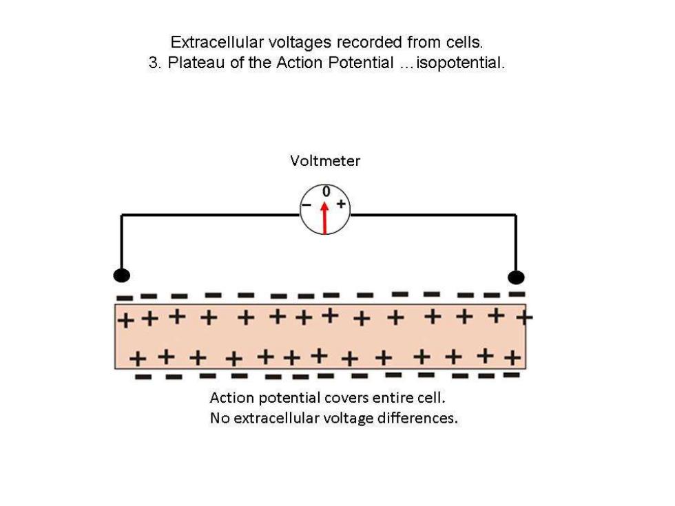
Extracellular voltages recorded from cells. 3.Plateau of the Action Potential...isopotential. Voltmeter 0 ■=■=一■■==■■一 ++十++++++++++ +++++++十+++++ Action potential covers entire cell. No extracellular voltage differences
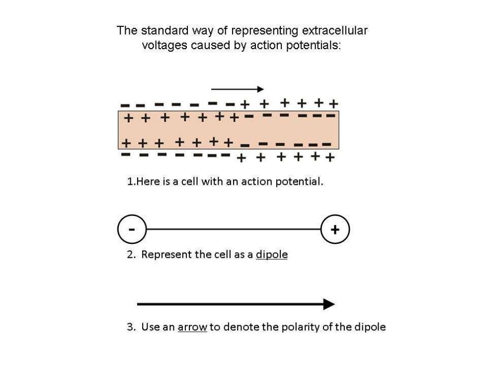
The standard way of representing extracellular voltages caused by action potentials: ▣=一=▣■▣++十+十十 十++十十+十=一一一一▣ 十++十十十+▣-■✉= ■■■■=■+十++十十 1.Here is a cell with an action potential. X 2.Represent the cell as a dipole 3.Use an arrow to denote the polarity of the dipole