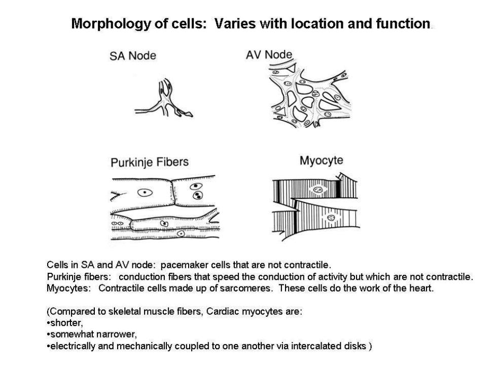
Morphology of cells:Varies with location and function SA Node AV Node Purkinje Fibers Myocyte Cells in SA and AV node:pacemaker cells that are not contractile. Purkinje fibers:conduction fibers that speed the conduction of activity but which are not contractile. Myocytes:Contractile cells made up of sarcomeres.These cells do the work of the heart. (Compared to skeletal muscle fibers,Cardiac myocytes are: .shorter, .somewhat narrower. .electrically and mechanically coupled to one another via intercalated disks

The heart is a syncytium.Action potentials are conducted from muscle cell to muscle cell in the heart.Unlike skeletal muscle,no nerves are involved in conduction of activity through the heart.Cells are electrically coupled to each other through intercalated discs,which contain gap junction channels Intercalated disc Gap junction (nexus) they bifurcate:each cell is Y-shaped,and contacts more than one downstream myocyte
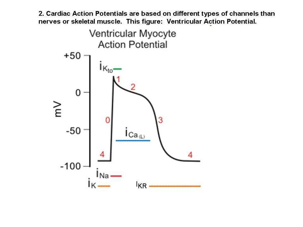
2.Cardiac Action Potentials are based on different types of channels than nerves or skeletal muscle.This figure:Ventricular Action Potential. Ventricular Myocyte Action Potential +50 0 金 0 3 -50 ICa 4 4 -100 INa lKR
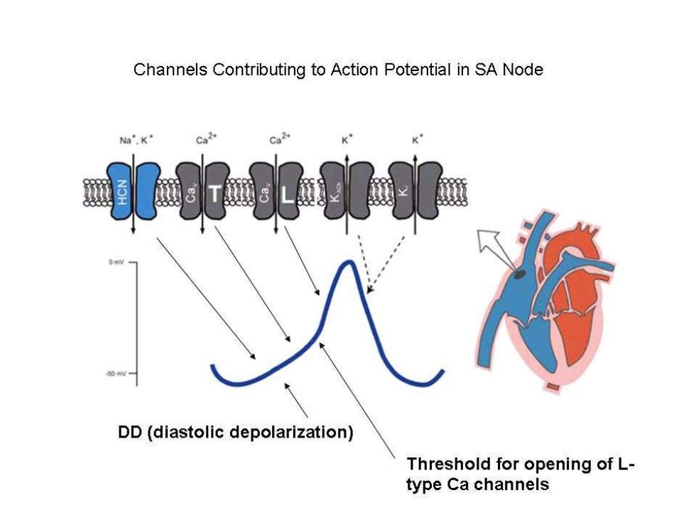
Channels Contributing to Action Potential in SA Node Na".K" Co 2. K DD(diastolic depolarization) Threshold for opening of L- type Ca channels
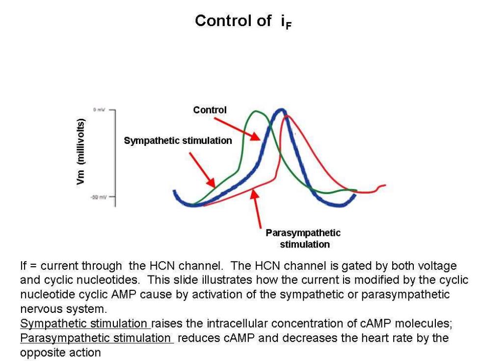
Control of i Control Sympathetic stimulation Parasympathetic stimulation If current through the HCN channel.The HCN channel is gated by both voltage and cyclic nucleotides.This slide illustrates how the current is modified by the cyclic nucleotide cyclic AMP cause by activation of the sympathetic or parasympathetic nervous system. Sympathetic stimulation raises the intracellular concentration of cAMP molecules; Parasympathetic stimulation reduces cAMP and decreases the heart rate by the opposite action