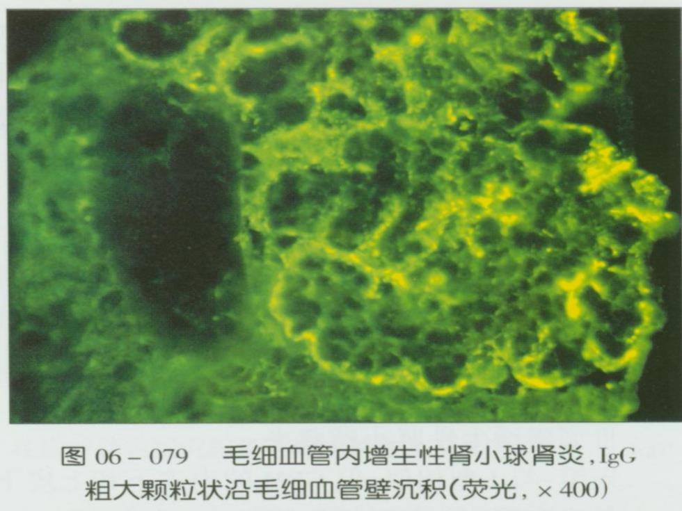
图06-079毛细血管内增生性肾小球肾炎,IgG 粗大颗粒状沿毛细血管壁沉积(荧光,×400)

Electron micrograph of a normal glomerulus Urinary space Electron micrograph of a normal glomerular capillary loop showing the fenestrated endothelial cell(Endo),the glomerular basement membrane(GBM),and the epithelial cells with its interdigitating foot processes(arrow).The GBM is thin,and no electron-dense deposits are present.Two normal platelets are seen in the capillary lumen. Platele Electron micrograph of postinfectious glomerulonephritis Urinary Electron micrograph shows subepithelial deposits(D)with a space semilunar,hump-shaped appearance in postinfectious glomerulonephritis.The humps sit on top of the glomerular basement membrane (GBM).A neutrophil is attached to the denuded GBM,contributing to the glomerular inflammation. Neutrophil attraction requires the initial presence of subepithelial immune deposits so that complement chemoattractants have access to the systemic circulation

CLINICAL PRESENTATION o Asymptomatic,microscopic hematuria o An antecedent history of a Gas skin or throat infection between 1-3 weeks following GAS pharyngitis between 3-6 weeks following GAS skin infection o Full-blown acute nephritic syndrome急性肾炎综合征 。Red to brown urine Proteinuria(can reach the nephrotic range) 。Edema ·Hypertension An elevation in serum creatinine
CLINICAL PRESENTATION Asymptomatic, microscopic hematuria An antecedent history of a GAS skin or throat infection between 1-3 weeks following GAS pharyngitis between 3-6 weeks following GAS skin infection Full-blown acute nephritic syndrome 急性肾炎综合征 Red to brown urine Proteinuria (can reach the nephrotic range) Edema Hypertension An elevation in serum creatinine
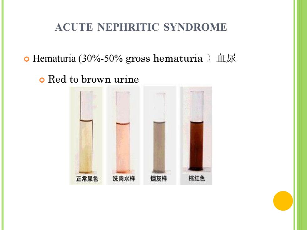
ACUTE NEPHRITIC SYNDROME o Hematuria(30%-50%gross hematuria)血尿 o Red to brown urine 正常尿色 洗肉水样 烟灰样 棕红色
ACUTE NEPHRITIC SYNDROME Hematuria (30%-50% gross hematuria )血尿 Red to brown urine

ACUTE NEPHRITIC SYNDROME o Proteinuria(can reach the nephrotic rangel)蛋白尿
ACUTE NEPHRITIC SYNDROME Proteinuria (can reach the nephrotic range) 蛋白尿