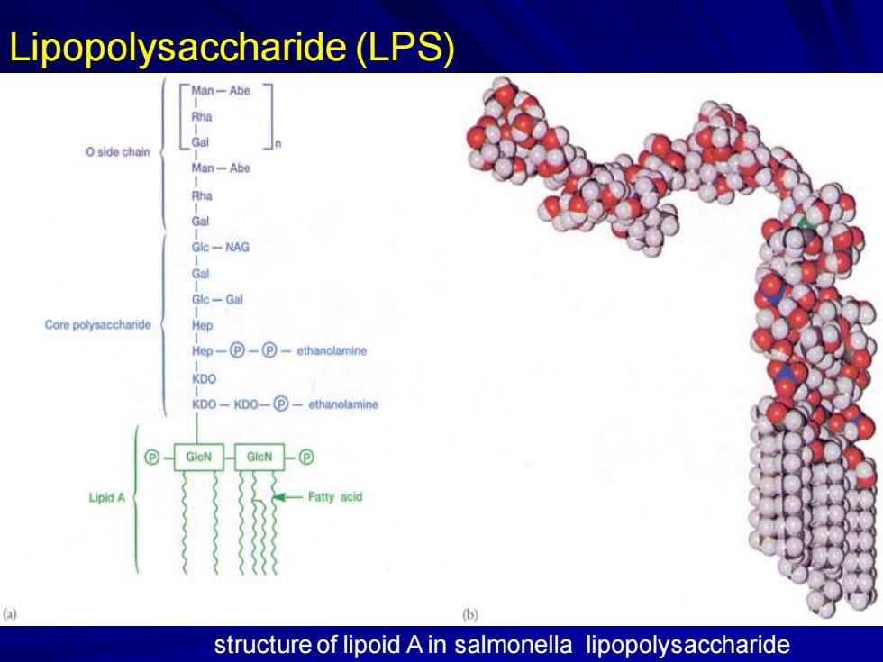
Lipopolysaccharide(LPS) Man-Abe Gal O side chain Man-Abe Rha Gal Gle-NAG Gal Glc-Gal Core polysaccharide Hep Hep-⊙-包-ethanolamine KDO KDO-KDO-P-ethanolamine ⊙ GleN GlcN Lipid A Fatty acid a structure of lipoid A in salmonella lipopolysaccharide
Lipopolysaccharide (LPS) structure of lipoid A in salmonella lipopolysaccharide
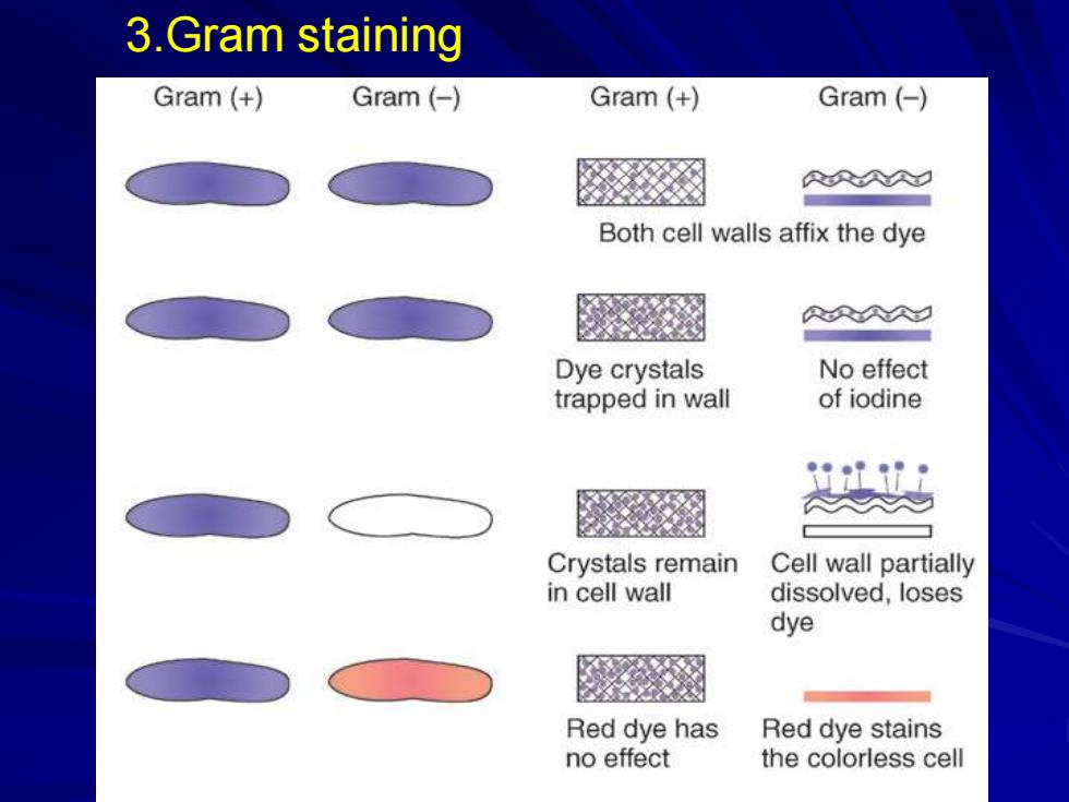
3.Gram staining Gram (+ Gram(-) Gram (+ Gram (- RRA Both cell walls affix the dye 刻 AAA久☑ Dye crystals No effect trapped in wall of iodine 妈 Crystals remain Cell wall partially in cell wall dissolved,loses dye Red dye has Red dye stains no effect the colorless cell
3.Gram staining
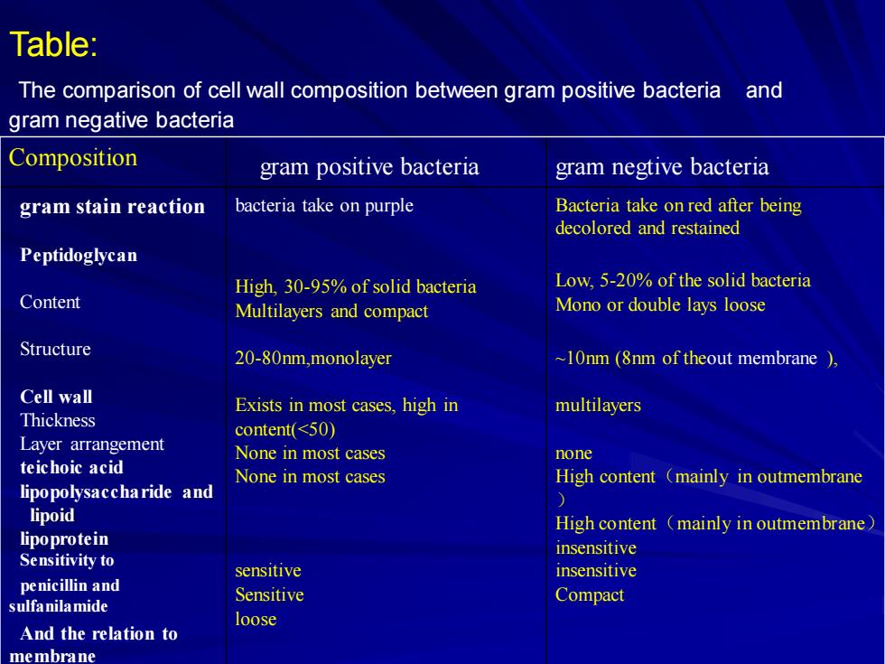
Table: The comparison of cell wall composition between gram positive bacteria and gram negative bacteria Composition gram positive bacteria gram negtive bacteria gram stain reaction bacteria take on purple Bacteria take on red after being decolored and restained Peptidoglycan High,30-95%of solid bacteria Low,5-20%of the solid bacteria Content Multilayers and compact Mono or double lays loose Structure 20-80nm,monolayer ~10nm(8nm of theout membrane ) Cell wall Exists in most cases,high in multilayers Thickness content(<50) Layer arrangement None in most cases none teichoic acid None in most cases High content (mainly in outmembrane lipopolysaccharide and lipoid High content (mainly in outmembrane) lipoprotein insensitive Sensitivity to sensitive insensitive penicillin and Sensitive sulfanilamide Compact loose And the relation to membrane
Composition gram positive bacteria gram negtive bacteria gram stain reaction Peptidoglycan Content Structure Cell wall Thickness Layer arrangement teichoic acid lipopolysaccharide and lipoid lipoprotein Sensitivity to penicillin and sulfanilamide And the relation to membrane bacteria take on purple High, 30-95% of solid bacteria Multilayers and compact 20-80nm,monolayer Exists in most cases, high in content(<50) None in most cases None in most cases sensitive Sensitive loose Bacteria take on red after being decolored and restained Low, 5-20% of the solid bacteria Mono or double lays loose ~10nm (8nm of theout membrane ), multilayers none High content(mainly in outmembrane ) High content(mainly in outmembrane) insensitive insensitive Compact Table: The comparison of cell wall composition between gram positive bacteria and gram negative bacteria
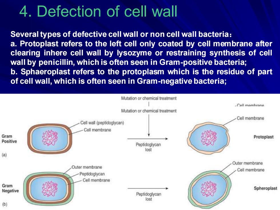
4.Defection of cell wall Several types of defective cell wall or non cell wall bacteria: a.Protoplast refers to the left cell only coated by cell membrane after clearing inhere cell wall by lysozyme or restraining synthesis of cell wall by penicillin,which is often seen in Gram-positive bacteria; b.Sphaeroplast refers to the protoplasm which is the residue of part of cell wall,which is often seen in Gram-negative bacteria; Mutation or chemical treatment Call momhrane Mutation or chemical treatment Cell membrane Cell wall (peptidoglycan) Cell membrane Gram Protoplast Positive Peptidoglycan lost (a) Outer membrane Outer membrane Cell membrane Peptidoglycan Cell membrane Gram Negative Spheroplast Peptidoglycan lost )
4. Defection of cell wall Several types of defective cell wall or non cell wall bacteria: a. Protoplast refers to the left cell only coated by cell membrane after clearing inhere cell wall by lysozyme or restraining synthesis of cell wall by penicillin, which is often seen in Gram-positive bacteria; b. Sphaeroplast refers to the protoplasm which is the residue of part of cell wall, which is often seen in Gram-negativebacteria;
3L-form Bacteria:L-form Bacteria special refer to transmissibility stable cell wall deficient strains, shaped by its natural mutant in the laboratory.In 1935,Lister Research Institute of preventive Medicine in England found out a kind of cell wall deficient bacteria,formed by natural mutant.- Streptobacillus moniliformis.it cell is big,and it is sensitive to the osmotic pressure,engendering small colonies in the solid culture media,like omelette .because of the first letter of Lister Research Institute is“L”,so named L-form Bacteria
③L-form Bacteria: L-form Bacteria special refer to transmissibility stable cell wall deficient strains, shaped by its natural mutant in the laboratory. In 1935, Lister Research Institute of preventive Medicine in England found out a kind of cell wall deficient bacteria, formed by natural mutant.- Streptobacillus moniliformis. it cell is big, and it is sensitive to the osmotic pressure, engendering small colonies in the solid culture media, like omelette .because of the first letter of Lister Research Institute is “L”, so named L-form Bacteria