MORPHOLOGY OF NECROSIS 1。 Nuclear changes:karyolysis,pyknosis karyorrhexis 2. Cytoplasmic changes:increased eosinophilia 3. Fates of necrotic cells:Dead cells may be replaced by myelin figures,which are further degraded into fatty acids binding calcium salts
MORPHOLOGY OF NECROSIS 1. Nuclear changes: karyolysis, pyknosis, karyorrhexis 2. Cytoplasmic changes: increased eosinophilia 3. Fates of necrotic cells: Dead cells may be replaced by myelin figures, which are further degraded into fatty acids binding calcium salts

Patterns of Tissue Necrosis o Coagulative necrosis o Liquefactive necrosis Gangrenous necrosis O Caseous necrosis O Fat necrosis O Fibrinoid necrosis
Patterns of Tissue Necrosis Coagulative necrosis Liquefactive necrosis Gangrenous necrosis Caseous necrosis Fat necrosis Fibrinoid necrosis
Coagulative necrosis A wedge-shaped kidney infarct (yellow)with preservation of the outlines. Microscopic view of the edge of the infarct,with normal kidney (N) and necrotic cells in the infarct (I).The necrotic cells show preserved outlines with loss of nuclei,and an inflammatory infiltrate is present (difficult to discern at this magnification)
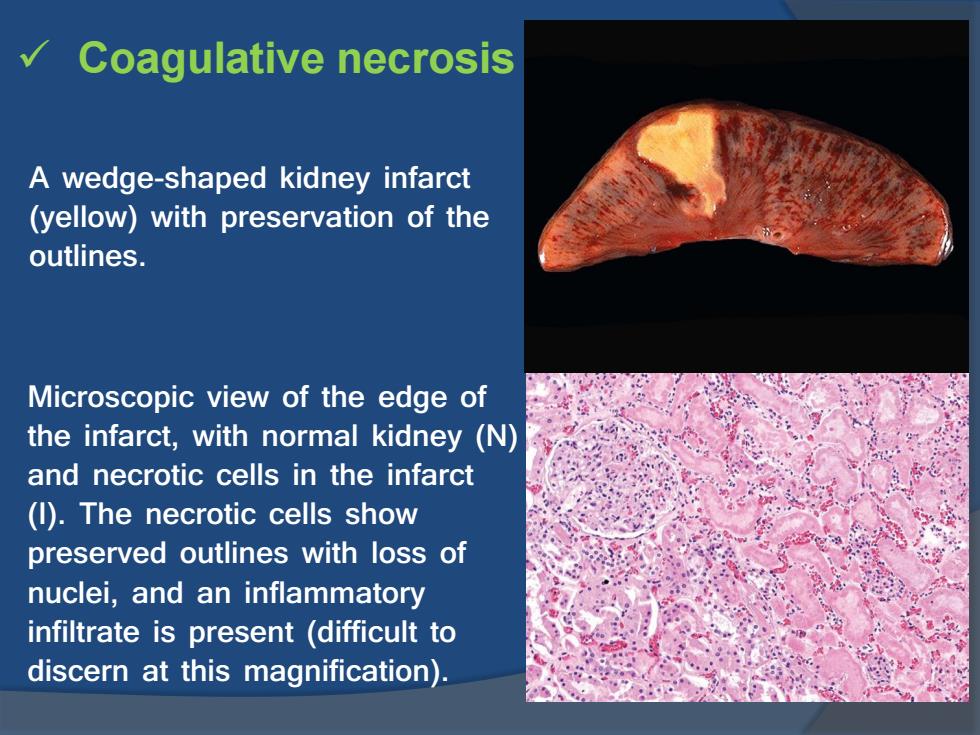
Coagulative necrosis A wedge-shaped kidney infarct (yellow) with preservation of the outlines. Microscopic view of the edge of the infarct, with normal kidney (N) and necrotic cells in the infarct (I). The necrotic cells show preserved outlines with loss of nuclei, and an inflammatory infiltrate is present (difficult to discern at this magnification)

Coagulative necrosis in cardiac infarction
Coagulative necrosis in cardiac infarction

Liquefactive necrosis An infarct in the brain showing dissolution of the tissue. The cystic space contains necrotic cell debris and macrophages filled with phagocytosed material
Liquefactive necrosis An infarct in the brain showing dissolution of the tissue. The cystic space contains necrotic cell debris and macrophages filled with phagocytosed material