Week 1 Quiz 1 Pathology Cell Injury Two days ago,you stubbed your big toe while hurrying to the gym. Now there is an ecchymosis unde r the nail bed as shown above.Which of the following cytologic features is associated with this reversible cell injury? A.Apoptosis B.Liquefactive necrosis C.Lysosome rupture D.Nuclear pyknosis E.Swelling of the mitochondria Ans:E ired leading to accumulation of intracellular scecellular volne resomssottase grad ions.The s with the mito drial me control.lea death: h ng to swelling.Apoptosis is th is process 1. process of enzymatic degradation and protein denaturation in the brain in reaction to exogenous injury (infarct or infection).Lysosome rupture and nuclear pyknosis are cellular changes of necrosis that occur in reaction to irreversible cell iniury. 2.A 16 year-old boy sustained blunt trauma to the abdomen when the vehicle he was driving struck a bridge abutment at high speed.Peritoneal lavage(washing the peritoneal cavity with saline)showed a hemoperitoneum(blood in the abdominal cavity),and at laparotomy(surgical exploration of the abdomen),a small portion of the left lobe of the liver was removed because of -1
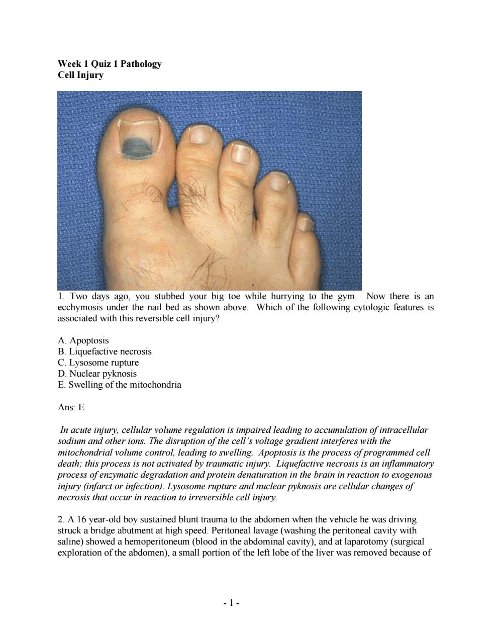
- 1 - Week 1 Quiz 1 Pathology Cell Injury 1. Two days ago, you stubbed your big toe while hurrying to the gym. Now there is an ecchymosis under the nail bed as shown above. Which of the following cytologic features is associated with this reversible cell injury? A. Apoptosis B. Liquefactive necrosis C. Lysosome rupture D. Nuclear pyknosis E. Swelling of the mitochondria Ans: E In acute injury, cellular volume regulation is impaired leading to accumulation of intracellular sodium and other ions. The disruption of the cell’s voltage gradient interferes with the mitochondrial volume control, leading to swelling. Apoptosis is the process of programmed cell death; this process is not activated by traumatic injury. Liquefactive necrosis is an inflammatory process of enzymatic degradation and protein denaturation in the brain in reaction to exogenous injury (infarct or infection). Lysosome rupture and nuclear pyknosis are cellular changes of necrosis that occur in reaction to irreversible cell injury. 2. A 16 year-old boy sustained blunt trauma to the abdomen when the vehicle he was driving struck a bridge abutment at high speed. Peritoneal lavage (washing the peritoneal cavity with saline) showed a hemoperitoneum (blood in the abdominal cavity), and at laparotomy (surgical exploration of the abdomen), a small portion of the left lobe of the liver was removed because of
the injury.Several weeks later,a CT scan of the abdomen shows that the liver is nearly normal in size again.Which of the following processes best explains this phenomenon? A.Apoptosis B.Dysplasia C.Fatty change D.Hydropic change E.Hyperplasia Ans:E in the mber of cells.which in this case is ar enter l grow py.Apoptosis is shape,and size.Fatty change (abnormal accumulation of lipids in parenchyma cells)usually occurs in the liver.In the heart,it would occur in the myocardium,not on the valves.Hydropic change is acute.reversible cell swelling in response to injury. 3.On a routine visit to the physician,an otherwise healthy 51 year-old man has an elevated blood pressure of 150/95 mm Hg.If the patient's hypertension remains untreated for years, which of the following cellular alterations will most likely be seen in the myocardium(heart muscle)? A.Atrophy B.Hemosiderosis C.Hypertrophy D.Metaplasia E Sclerosis Ans:C places increased stress on the lejt ventricle.which msi increase contractlejorce a pres. not possible. Instea cells in number or size of cells due to decreased stimulation or stress. an iron overload disorder unrelated to hypertension.Metaplasia is replacement ofone cell type with another due to irritation or environmental exposure.Sclerosis describes a hardening ofa structure,often due to replacement with connective tissue. -2
- 2 - the injury. Several weeks later, a CT scan of the abdomen shows that the liver is nearly normal in size again. Which of the following processes best explains this phenomenon? A. Apoptosis B. Dysplasia C. Fatty change D. Hydropic change E. Hyperplasia Ans: E Hyperplasia describes the increase in the number of cells, which in this case is a response to injury. It requires stimulation of cell sin G0 to enter the cell cycle and multiply. Apoptosis is programmed cell death. Dysplasia refers to abnormal growth of cells and loss of proper orientation, shape, and size. Fatty change (abnormal accumulation of lipids in parenchymal cells) usually occurs in the liver. In the heart, it would occur in the myocardium, not on the valves. Hydropic change is acute, reversible cell swelling in response to injury. 3. On a routine visit to the physician, an otherwise healthy 51 year-old man has an elevated blood pressure of 150/95 mm Hg. If the patient’s hypertension remains untreated for years, which of the following cellular alterations will most likely be seen in the myocardium (heart muscle)? A. Atrophy B. Hemosiderosis C. Hypertrophy D. Metaplasia E. Sclerosis Ans: C Hypertension places increased stress on the left ventricle, which must increase contractile force to eject blood against increased pressure. Cardiac tissue is terminally differentiated, thus hyperplasia is not possible. Instead, the cells increase in size (hypertrophy). Atrophy is a reduction in the number or size of cells due to decreased stimulation or stress. Hemosiderosis is an iron overload disorder unrelated to hypertension. Metaplasia is replacement of one cell type with another due to irritation or environmental exposure. Sclerosis describes a hardening of a structure, often due to replacement with connective tissue

©Elsevier2005 4.A 72 vear-old man died suddenly and unexpectedly from congestive heart failure.At autopsy the heart was very enlarged and weighed 580 grams.There was marked left ventricular hypertrophy and minimal oro arterial atherosclerosis.A serum chemistry panel ordered ath sho malities.Which of the followi ing the figure processes best accounts for the appearance of his aortic valve,which is seen Amyloidosis Dystrophic Fatty Ch ange D.Hemosiderosis E.Lipofuscin deposition Ans:B Dystrophic calcification is an extracellular deposition of calcium in the setting of persistent necrotic tissue.The figure shows calcium deposits on the aortic valve leaflets that impair blood flow,causing left ventricular hypertrophy.Amyloidosis is a large category of disorders characterized by diverse extracellular protein denosits not visible to the naked eve fatty chang (abnormal accumulation of lipids in parenchymal cells)usually occurs in the liver.In the heart, it would occur in the mvocardium,not on the valves.Hemosiderosis is an iron overload disorder that does not cause extracellular calcium deposition.Lipofuscin is the result of accumulated oxidized and cross-linked proteins:these depo sits are not visible to the naked eye. -3
- 3 - 4. A 72 year-old man died suddenly and unexpectedly from congestive heart failure. At autopsy, the heart was very enlarged and weighed 580 grams. There was marked left ventricular hypertrophy and minimal coronary arterial atherosclerosis. A serum chemistry panel ordered prior to death showed no abnormalities. Which of the following pathologic processes best accounts for the appearance of his aortic valve, which is seen in the figure? A. Amyloidosis B. Dystrophic calcification C. Fatty Change D. Hemosiderosis E. Lipofuscin deposition Ans: B Dystrophic calcification is an extracellular deposition of calcium in the setting of persistent necrotic tissue. The figure shows calcium deposits on the aortic valve leaflets that impair blood flow, causing left ventricular hypertrophy. Amyloidosis is a large category of disorders characterized by diverse extracellular protein deposits not visible to the naked eye. Fatty change (abnormal accumulation of lipids in parenchymal cells) usually occurs in the liver. In the heart, it would occur in the myocardium, not on the valves. Hemosiderosis is an iron overload disorder that does not cause extracellular calcium deposition. Lipofuscin is the result of accumulated oxidized and cross-linked proteins; these deposits are not visible to the naked eye

©Elsevier2005 5.A38 year-old woman experienced severe abdominal pain with hypotension and shock that led to her death within 36 hours after the onset of the pain.An autopsy was performed.The mesentery is shown above.The fatty tissue is covered with multiple yellow areas of tissue injury. Which of the following events has most likely occurred? A.Acute pancreatitis B.Gangrenous cholecystitis C.Hepatitis B virus infection D.Small intestinal infarction E.Tuberculous lymphadenitis Ans:A e pancreatitis which causes en chalk matic fat ne the would sh n by ecro organ ns. clinical features would have more green-bioo ikely b RUO pain an orexia,but no no association of pancreatitis and Hepatitis B infection.although case reports have suggested an increased mortality in the event of co-infection.Small bowel infarction would demonstrate dusky serosa and hemorrhage,not chalky-white nodules.Lymphadenitis caused by tuberculosis is most commonly of the cervical nodes,but infection of peritoneal,peri-pancreatic,mesenteric,or hepatic lymph nodes is possible.These infections are unlikely to lead to rapidly progressive shock and death.For example,hepatic nodal involvement might cause slow onset of jaundice and portal hypertension. -4
- 4 - 5. A 38 year-old woman experienced severe abdominal pain with hypotension and shock that led to her death within 36 hours after the onset of the pain. An autopsy was performed. The mesentery is shown above. The fatty tissue is covered with multiple yellow areas of tissue injury. Which of the following events has most likely occurred? A. Acute pancreatitis B. Gangrenous cholecystitis C. Hepatitis B virus infection D. Small intestinal infarction E. Tuberculous lymphadenitis Ans: A These symptoms are classic for acute pancreatitis which causes enzymatic fat necrosis of the peri-pancreatic or abdominal fat via lipase, as shown by the chalky-white nodules above. Gangrenous cholecystitis would show a green-black necrotic organ with some perforations; clinical features would have more likely been RUQ pain and anorexia, but not shock. There is no association of pancreatitis and Hepatitis B infection, although case reports have suggested an increased mortality in the event of co-infection. Small bowel infarction would demonstrate dusky serosa and hemorrhage, not chalky-white nodules. Lymphadenitis caused by tuberculosis is most commonly of the cervical nodes, but infection of peritoneal, peri-pancreatic, mesenteric, or hepatic lymph nodes is possible. These infections are unlikely to lead to rapidly progressive shock and death. For example, hepatic nodal involvement might cause slow onset of jaundice and portal hypertension

6.In an experiment,cells are subjected to radiant energy in the form of x-rays This results in cell injury caused by hydrolysis of water and free radical generation.Which of the following cellular enzymes normally protects cells from free radical injury? A.Endonuclease B.Glutathione peroxidase C.Lactate dehydrogenase D.Phosholipase E.Protease Ans:B Glutathione peroxidase is one of three key enzymes that neutralize free radicals (the others are superoxide dismuase and caalase).Endonuclease cleaves phosphodiester bonds chain of tides. Lactate dehydrogend 2 the re nate to e and the fatty acids. e bonds in the process ofproteolysis robzes lipids 7.A30 year old man fract tures his left femur in a biking a cide d his leg is pl ced in a plaster cast. fter the leg has been immobilized for several week the diameter the left cal This change is most likely to result from which of the following alterations in the calf muscles A.Aplasia B.Atrophy C.Dystrophy D.Hypoplasia E.Metaplasia Ans:B Atrophy refers to the reduction in n umber or size ofcells.In this case,disuse of the limb results d neuronal stimulation to the skeletal n Aplasia is the failr of his leg. which leads to atre etof元 m D the dege of tissue Hypop acement ofone cell type with another in response to stress or irritation 8. A experiment ana yme activity associated wit A.Endothelial cells B.Erythrocytes C.Germ cells D.Neurons E.Neutrophils 5
- 5 - 6. In an experiment, cells are subjected to radiant energy in the form of x-rays. This results in cell injury caused by hydrolysis of water and free radical generation. Which of the following cellular enzymes normally protects cells from free radical injury? A. Endonuclease B. Glutathione peroxidase C. Lactate dehydrogenase D. Phosholipase E. Protease Ans: B Glutathione peroxidase is one of three key enzymes that neutralize free radicals (the others are superoxide dismutase and catalase). Endonuclease cleaves phosphodiester bonds of a chain of nucleotides. Lactate dehydrogenase catalyzes the reduction of pyruvate to lactate and the oxidation of lactate to pyruvate in the liver (Cori cycle). Phospholipase hydrolyzes lipids into fatty acids. Protease hydrolyzes peptide bonds in the process of proteolysis. 7. A 30 year-old man fractures his left femur in a biking accident, and his leg is placed in a plaster cast. After the leg has been immobilized for several weeks, the diameter of the left calf has decreased. This change is most likely to result from which of the following alterations in the calf muscles? A. Aplasia B. Atrophy C. Dystrophy D. Hypoplasia E. Metaplasia Ans: B Atrophy refers to the reduction in number or size of cells. In this case, disuse of the limb results in decreased neuronal stimulation to the skeletal muscles of his leg, which leads to atrophy. Aplasia is the failure of a structure to form. Dystrophy is the degeneration of tissue due to a pathologic process. Hypoplasia refers to incomplete development of a tissue or organ. Metaplasia is replacement of one cell type with another in response to stress or irritation. 8. An experiment analyzes cells for enzyme activity associated with sustained cellular proliferation. Which of the following cell types is most likely to have the highest telomerase activity? A. Endothelial cells B. Erythrocytes C. Germ cells D. Neurons E. Neutrophils