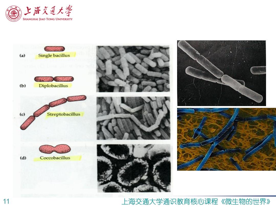
上游充通大粤 SHANGHAI JIAO TONG UNIVERSITY (a) Single bacillus @ (b) Diplobacillus (c) Streptobacillus (d) Coccobacillus 11 上海交通大学通识教育核心课程 《微生物的世界》
11 上海交通大学通识教育核心课程《微生物的世界》
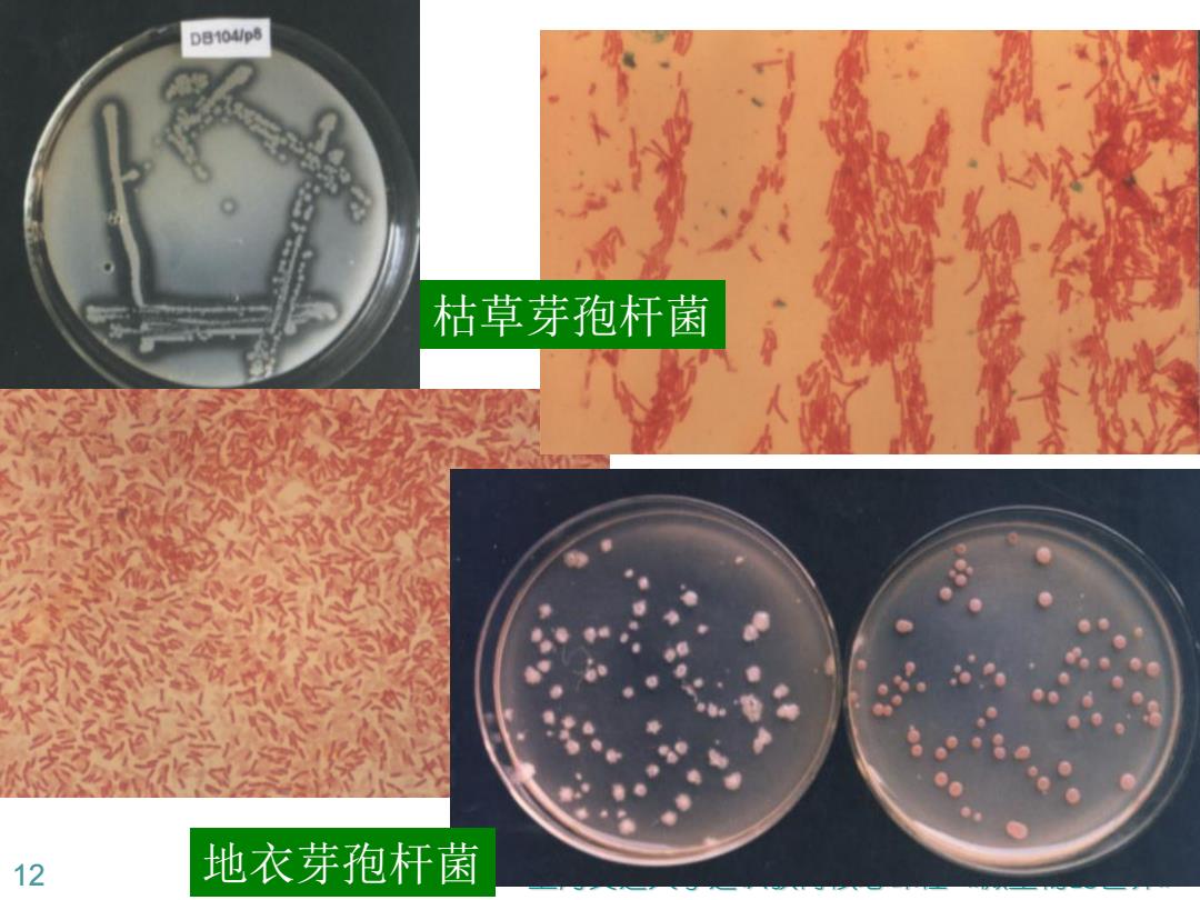
DB104/p8 枯草芽孢杆菌 12 地衣芽孢杆菌
12 上海交通大学通识教育核心课程《微生物的世界》 枯草芽孢杆菌 地衣芽孢杆菌
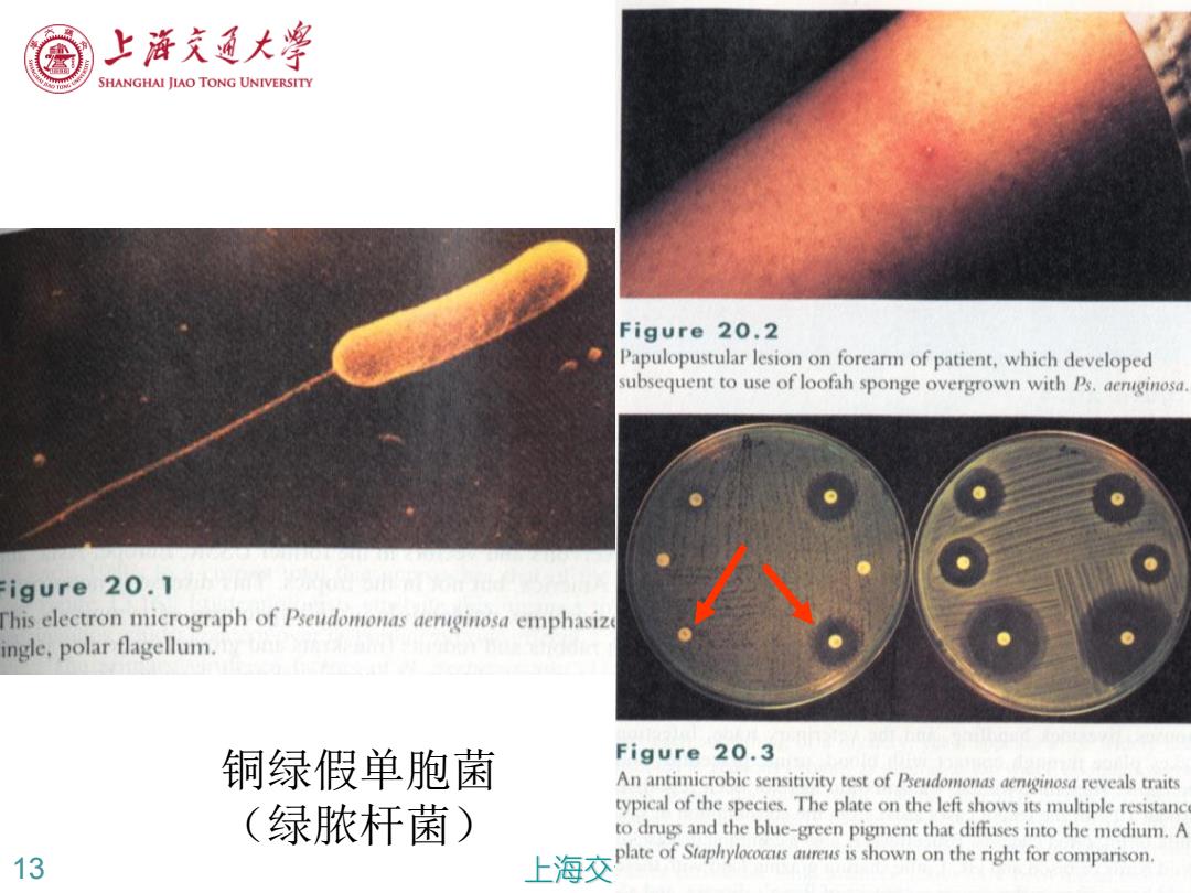
上帝充通大粤 SHANGHAI JIAO TONG UNIVERSITY Figure 20.2 Papulopustular lesion on forearm of patient,which developed subsequent to use of loofah sponge overgrown with Ps.aenginosa. igure 20.1 his electron micrograph of Pseudomonas aerginosa emphasize ingle,polar flagellum. 铜绿假单胞菌 Figure 20.3 An antimicrobic sensitivity test of Pseudomonas aenginosa reveals traits (绿脓杆菌) typical of the species.The plate on the left shows its multiple resistanc to drugs and the blue-green pigment that diffuses into the medium.A 13 上海交 plate of Staphrylococs aureus is shown on the right for comparison
13 上海交通大学通识教育核心课程 《微生物的世界 》 铜绿假单胞菌 (绿脓杆菌)

、.业上A1的 结核分枝杆菌 e19.20 escent acid-fast stain of M.tuberculosis from sputum.Smears uated in terms of the number of AFB seen per field.This y is then applied to a scale ranging from 0 to 4+,0 being no oserved and 4+being more than 9 AFB per field. 通大学通识教育核心课程 《微生物的世界》
14 上海交通大学通识教育核心课程《微生物的世界》 结核分枝杆菌
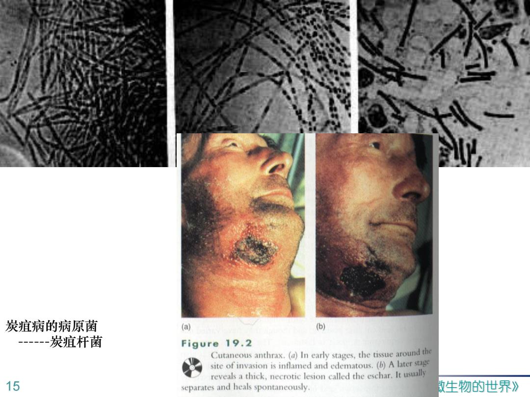
炭疽病的病原菌 (a) (b) --炭疽杆菌 Figure 19.2 Cutaneous anthrax.(a)In early stages,the tissue around the site of invasion is inflamed and edematous.(b)A later stage reveals a thick,necrotic lesion called the eschar.It usually 15 separates and heals spontaneously. 数生物的世界》
15 上海交通大学通识教育核心课程《微生物的世界》 炭疽病的病原菌 ------炭疽杆菌