Lewy Body At the left,an H and E stain demonstrates a rounded pink cytoplasmic Lewy body in a neuron of the Parkinson's disease. At the right,is an immunoperoxidase stain for better demonstration of the Lewy bodies
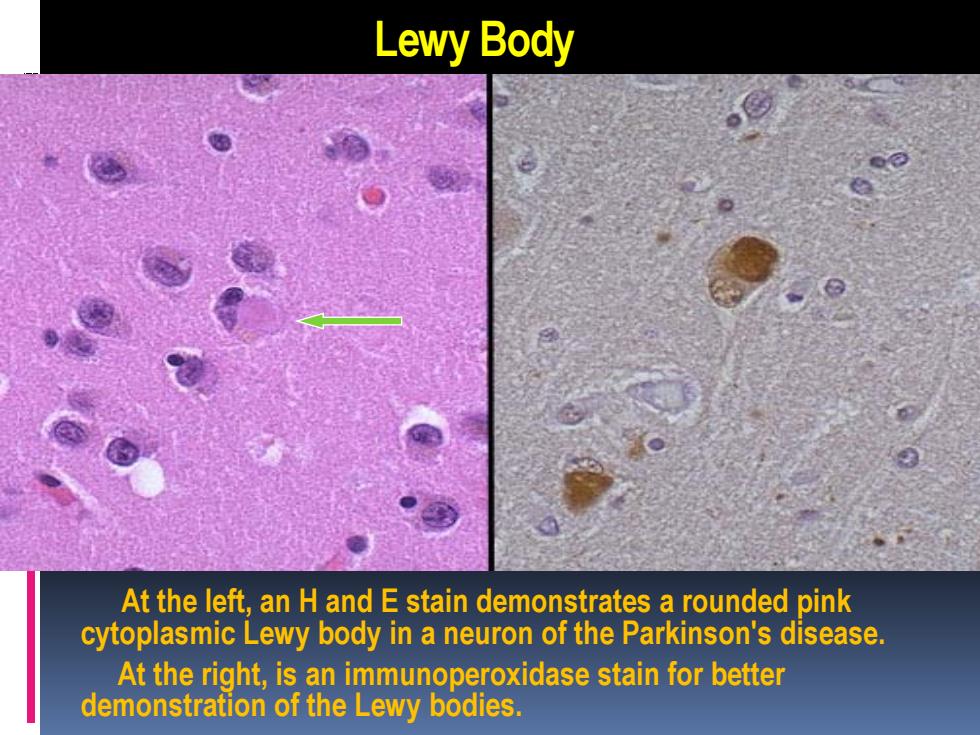
Lewy Body At the left, an H and E stain demonstrates a rounded pink cytoplasmic Lewy body in a neuron of the Parkinson's disease. At the right, is an immunoperoxidase stain for better demonstration of the Lewy bodies
Neurofibrillary Tangle I This is a neurofibrillary tangle of Alzheimer's disease.The tangle appears as long pink filaments in the cytoplasm. They are composed of cytoskeletal intermediate filaments
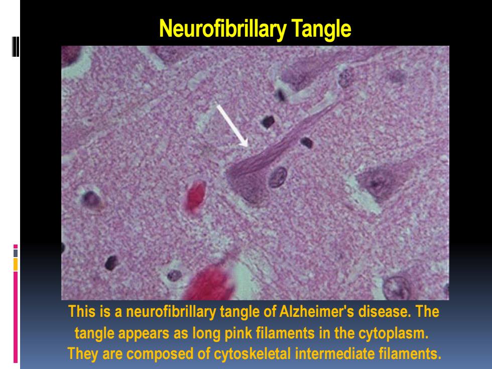
Neurofibrillary Tangle This is a neurofibrillary tangle of Alzheimer's disease. The tangle appears as long pink filaments in the cytoplasm. They are composed of cytoskeletal intermediate filaments

Neurofibrillary Tangle L Neurofibrillary tangles of Alzheimer's disease are also seen best with a silver stain,as shown here
Neurofibrillary Tangle Neurofibrillary tangles of Alzheimer's disease are also seen best with a silver stain, as shown here
Intranuclear Inclusion Cytomegalovirus(CMV)induces intranuclear inclusions, made prominent by the presence of clear halos
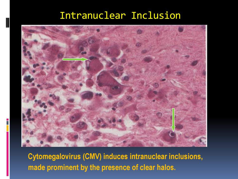
Intranuclear Inclusion Cytomegalovirus (CMV) induces intranuclear inclusions, made prominent by the presence of clear halos
Intracytoplasmic Inclusion Negri body.Rabies encephalitis is characterized by round, eosinophilic cytoplasmic inclusions that resemble an erythrocyte
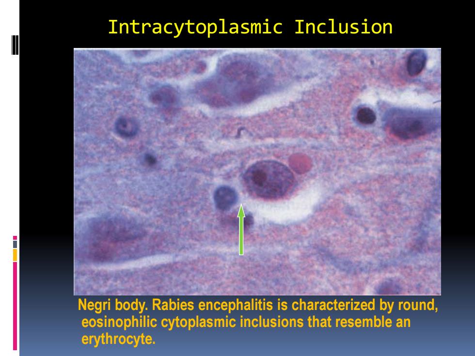
Intracytoplasmic Inclusion Negri body. Rabies encephalitis is characterized by round, eosinophilic cytoplasmic inclusions that resemble an erythrocyte