
Quadriceps femoris ■ Origin: Rectus femoris:anterior inferior iliac spine Vastus medialis:medial lip of linea aspera Vastus lateralis:lateral lip of linea aspera ▣Vastus intermedius: anterior surface of femur Insertion:tibial tuberosity via patellar ligament Action:extends leg at knee joint;rectus femoris also flexes thigh at hip joint Nerve supply:femoral n
Quadriceps femoris ◼ Origin: ❑ Rectus femoris: anterior inferior iliac spine ❑ Vastus medialis: medial lip of linea aspera ❑ Vastus lateralis: lateral lip of linea aspera ❑ Vastus intermedius: anterior surface of femur ◼ Insertion: tibial tuberosity via patellar ligament ◼ Action: extends leg at knee joint; rectus femoris also flexes thigh at hip joint ◼ Nerve supply: femoral n

Tibialis anterior Origin:lateral surface of tibia ■ Insertion:medial cuneiform and base of 1st metatarsal Action:dorsiflexes and inverts foot Nerve supply:deep peroneal n
Tibialis anterior ◼ Origin: lateral surface of tibia ◼ Insertion: medial cuneiform and base of 1st metatarsal ◼ Action: dorsiflexes and inverts foot ◼ Nerve supply: deep peroneal n
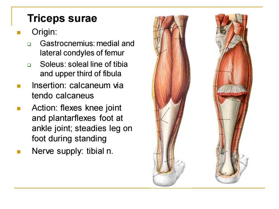
Triceps surae Origin: Gastrocnemius:medial and lateral condyles of femur Soleus:soleal line of tibia and upper third of fibula ■ Insertion:calcaneum via tendo calcaneus Action:flexes knee joint and plantarflexes foot at ankle joint;steadies leg on foot during standing Nerve supply:tibial n
Triceps surae ◼ Origin: ❑ Gastrocnemius: medial and lateral condyles of femur ❑ Soleus: soleal line of tibia and upper third of fibula ◼ Insertion: calcaneum via tendo calcaneus ◼ Action: flexes knee joint and plantarflexes foot at ankle joint; steadies leg on foot during standing ◼ Nerve supply: tibial n

Tibialis posterior Origin:posterior surface of tibia and ffibula and interosseous membrane Insertion:tuberosity of navicular,all cuniforms Action:plantarflexes and inverts foot Nerve supply:tibial n
Tibialis posterior ◼ Origin: posterior surface of tibia and ffibula and interosseous membrane ◼ Insertion: tuberosity of navicular, all cuniforms ◼ Action: plantarflexes and inverts foot ◼ Nerve supply: tibial n

Arteries of lower limb Femoral a. Continuation of the external iliac a. Begins midpoint of inguinal ligament Principal branch deep femeral a.股深动脉:arises from the posterolateral surface of the femoral artery about 5 cm below the inguinal ligament. Distributed to all three muscle compartments by medial and lateral femoral circumflex旋股内、外侧动脉 and four perforating arteries穿动脉 of deep femoral a
Arteries of lower limb Femoral a. ◼ Continuation of the external iliac a. ◼ Begins midpoint of inguinal ligament ◼ Principal branch deep femeral a.股深动脉: arises from the posterolateral surface of the femoral artery about 5 cm below the inguinal ligament. ◼ Distributed to all three muscle compartments by medial and lateral femoral circumflex旋股内、外侧动脉 and four perforating arteries 穿动脉 of deep femoral a