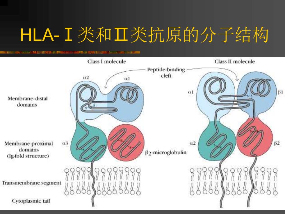
HLA-I类和Ⅱ类抗原的分子结构 重厚泽重通万■ Class I molecule Class II molecule Peptide-binding (2 cleft Membrane-distal domains Membrane-proximal domains B2-microglobulin (g-fold structure) 9997X99997999 999799990798 Transmembrane segment g6888888388 Cytoplasmic tail
HLA-Ⅰ类和Ⅱ类抗原的分子结构

(a) Peptide-binding (b) cleft al domain a1 domain a2 domain a helix B sheets a2 domain β2-microglobulin a3 domain LA-I类分子的三维空间构型图
HLA-Ⅰ类分子的三维空间构型图

(a) >HLA-I类分子抗原结合区的三维图
➢HLA-Ⅰ类分子抗原结合区的三维图
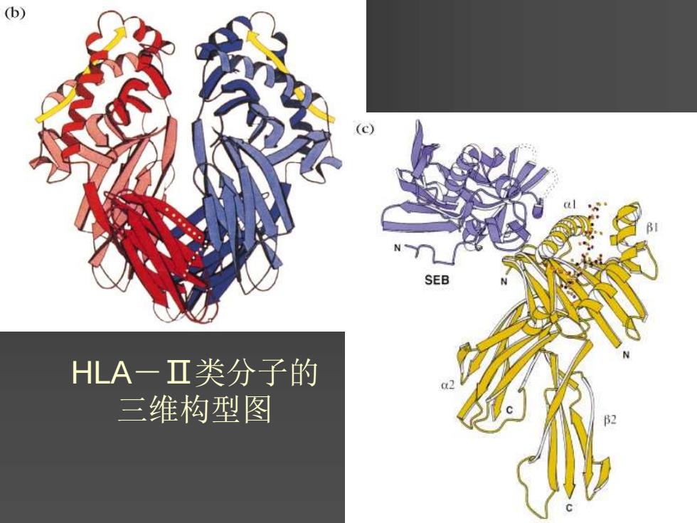
(b) N SEB HLA一Ⅱ类分子的 三维构型图
HLA-Ⅱ类分子的 三维构型图
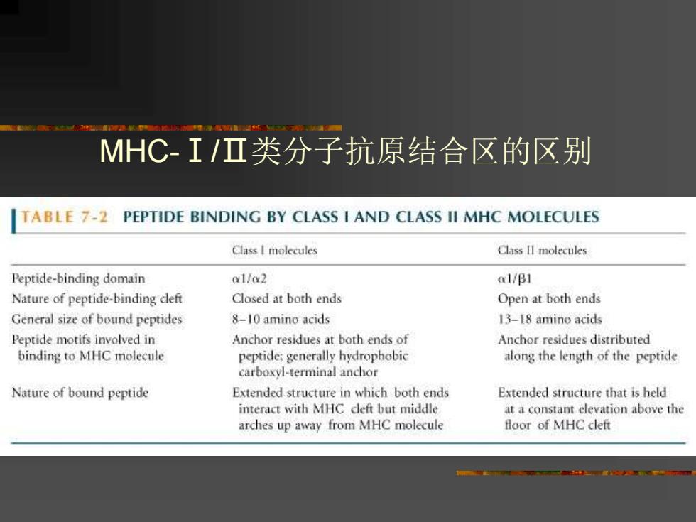
MHC-I/I类分子抗原结合区的区别 TABLE 7-2 PEPTIDE BINDING BY CLASS I AND CLASS II MHC MOLECULES Class I molecules Class II molecules Peptide-binding domain a1/x2 a1/B1 Nature of peptide-binding cleft Closed at both ends Open at both ends General size of bound peptides 8-10 amino acids 13-18 amino acids Peptide motifs involved in Anchor residues at both ends of Anchor residues distributed binding to MHC molecule peptide:generally hydrophobic along the length of the peptide carboxyl-terminal anchor Nature of bound peptide Extended structure in which both ends Extended structure that is held interact with MHC cleft but middle at a constant elevation above the arches up away from MHC molecule floor of MHC cleft
MHC-Ⅰ/Ⅱ类分子抗原结合区的区别