Low absorbance detected in samples .Check that samples were sufficiently induced and lysed(see Troubleshooting protein amplification page 9). .If clarified lysate is being tested,mix initial GST sample with 2X blocking buffer to give a final concentration of 1X blocking buffer. Poor day to day reproducibility between identical samples .Ensure that all incubation times are consistent.Reduction in GST capture incubation time can be reduced to >30 minutes with slightly reduced signal,but HRP/anti-GST conjugate incubation time can significantly reduce signal with every 15 minute decrease Alternative 4.Western blot analysis Anti-GST Antibody Wash buffer:.1%Tween 20in PBS(asabove) darynibecthe-Ty() 1.Separate the protein samples by SDS-PAGE. Although anti-GST antibody from Amersham Pharmacia Biotech has been cross-adsorbed with E.coli proteins,low levels of cross-reacting antibodies may remain.It is recommended always to run samples of E.coli sonicates that do not contain a recombinant pGEX plasmid and samples that contain the parental pGEX plasmid as controls. from the alectro o to anan Blocking of membrane 1.Transfer the which the proteins have been such asa Peri dish Add 50-200 ml of blocking/incubation buffer 3.Incubate for 1-16 hours at ambient temperature with gentle shaking. Longer incubation times with blocking buffer may reduce background signa 4.Decant and discard the buffer. 26
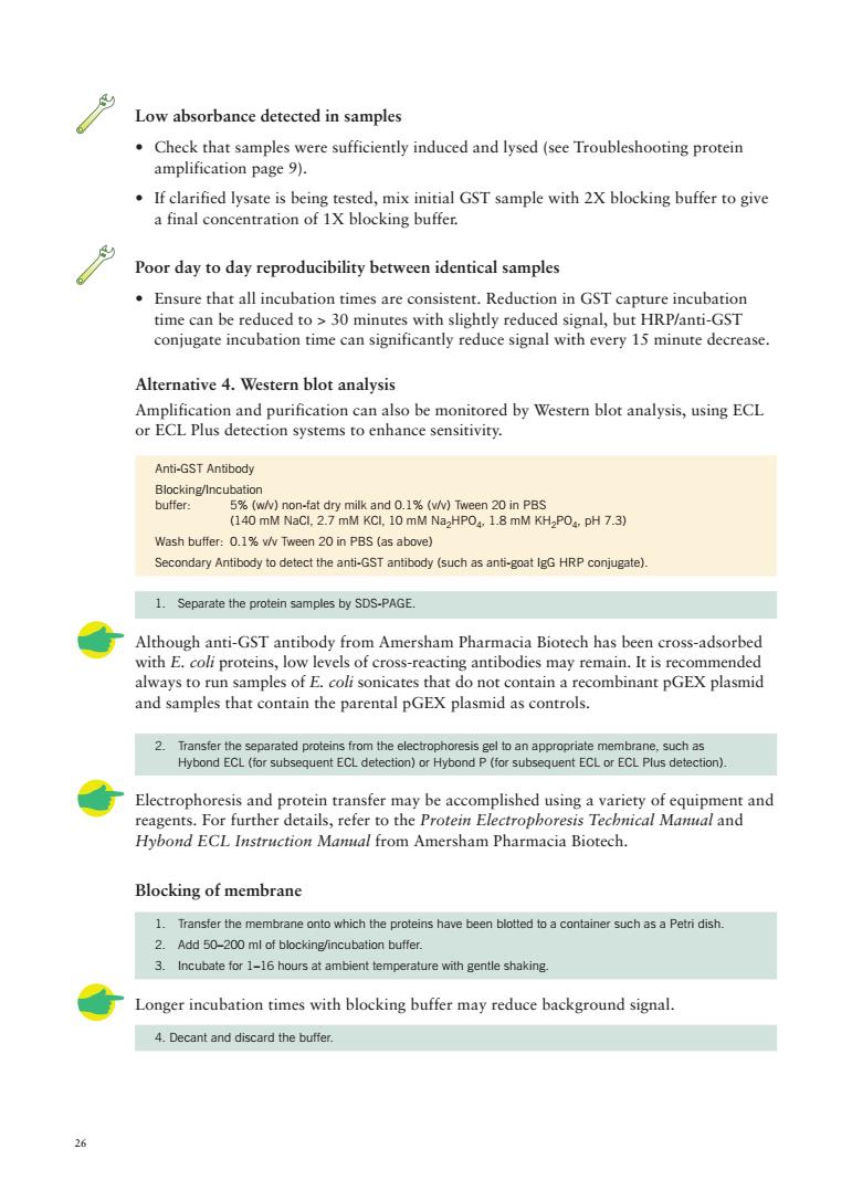
26 Anti-GST Antibody Blocking/Incubation buffer: 5% (w/v) non-fat dry milk and 0.1% (v/v) Tween 20 in PBS (140 mM NaCl, 2.7 mM KCl, 10 mM Na2HPO4, 1.8 mM KH2PO4, pH 7.3) Wash buffer: 0.1% v/v Tween 20 in PBS (as above) Secondary Antibody to detect the anti-GST antibody (such as anti-goat IgG HRP conjugate). 1. Separate the protein samples by SDS-PAGE. Although anti-GST antibody from Amersham Pharmacia Biotech has been cross-adsorbed with E. coli proteins, low levels of cross-reacting antibodies may remain. It is recommended always to run samples of E. coli sonicates that do not contain a recombinant pGEX plasmid and samples that contain the parental pGEX plasmid as controls. 2. Transfer the separated proteins from the electrophoresis gel to an appropriate membrane, such as Hybond ECL (for subsequent ECL detection) or Hybond P (for subsequent ECL or ECL Plus detection). Electrophoresis and protein transfer may be accomplished using a variety of equipment and reagents. For further details, refer to the Protein Electrophoresis Technical Manual and Hybond ECL Instruction Manual from Amersham Pharmacia Biotech. Blocking of membrane 1. Transfer the membrane onto which the proteins have been blotted to a container such as a Petri dish. 2. Add 50–200 ml of blocking/incubation buffer. 3. Incubate for 1–16 hours at ambient temperature with gentle shaking. Longer incubation times with blocking buffer may reduce background signal. 4. Decant and discard the buffer. Low absorbance detected in samples • Check that samples were sufficiently induced and lysed (see Troubleshooting protein amplification page 9). • If clarified lysate is being tested, mix initial GST sample with 2X blocking buffer to give a final concentration of 1X blocking buffer. Poor day to day reproducibility between identical samples • Ensure that all incubation times are consistent. Reduction in GST capture incubation time can be reduced to > 30 minutes with slightly reduced signal, but HRP/anti-GST conjugate incubation time can significantly reduce signal with every 15 minute decrease. Alternative 4. Western blot analysis Amplification and purification can also be monitored by Western blot analysis, using ECL or ECL Plus detection systems to enhance sensitivity

Anti-GST antibody nformation on optimization. 2.Pour the antibody-butfer mixture into the container with the membrane. 3.Incubate for 1 hour at ambient temperature with gentle shaking. 4.Decant and discard the antibody-buffer. 5.Rinse twice with 20-30 ml of blocking or wash buffer to remove most of the unbound antibody. 6.Decant and discard the ninses. 7.Wash the membrane with 20-30 ml of blocking or wash buffer for 10-60 minutes at ambient temperature with gentle shaking. 8.Discard the wash and repea Secondary antibody further information on optimization Pour the antibody-buffer mixture into the container with the membrane 3.Incubate for 1 hour at ambient temperature with gentle shaking. 4.Decant and discard the antibody-buffer. 5.Rinse twice with 20-30 ml of blocking or wash buffer to remove most of the unbound antibody. 6.Decant and discard the rinses. 7.Wash the membrane with 20-30ml of blockingor wash buffer for 1-60 minutes at ambient temperature Discard the wash and repea 9.Develop the blot with the appropriate substrate for the conjugated secondary antibody ECL and ECL Plus detection systems require very little antibody to achieve a sufficient the nt d se condary) ed in th h of a dy-buffe used by sc g down the protocol and perfo g the incubations in sealable plastic bags. Troubleshooting See also Purification and Detection Troubleshooting page 28. Multiple bands seen on Western blot analysis Anti-gST antibody from Amersham pharmacia biotech has been cross-absorbed against roteins and ted for its lack of pecific backgr hlot So e sources of the anti-GST antibody react with various E.coli proteins present in the fusion protein sample.Cross-adsorb the antibody with an E.coli sonicate to remove anti-E.coli antibodies.This E.coli must not contain the pGEX plasmid. 27
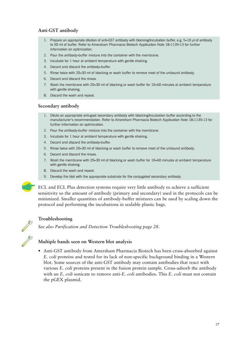
27 Anti-GST antibody 1. Prepare an appropriate dilution of anti-GST antibody with blocking/incubation buffer, e.g. 5–10 µl of antibody to 50 ml of buffer. Refer to Amersham Pharmacia Biotech Application Note 18-1139-13 for further information on optimization. 2. Pour the antibody-buffer mixture into the container with the membrane. 3. Incubate for 1 hour at ambient temperature with gentle shaking. 4. Decant and discard the antibody-buffer. 5. Rinse twice with 20–30 ml of blocking or wash buffer to remove most of the unbound antibody. 6. Decant and discard the rinses. 7. Wash the membrane with 20–30 ml of blocking or wash buffer for 10–60 minutes at ambient temperature with gentle shaking. 8. Discard the wash and repeat. Secondary antibody 1. Dilute an appropriate anti-goat secondary antibody with blocking/incubation buffer according to the manufacturer's recommendation. Refer to Amersham Pharmacia Biotech Application Note 18-1139-13 for further information on optimization. 2. Pour the antibody-buffer mixture into the container with the membrane. 3. Incubate for 1 hour at ambient temperature with gentle shaking. 4. Decant and discard the antibody-buffer. 5. Rinse twice with 20–30 ml of blocking or wash buffer to remove most of the unbound antibody. 6. Decant and discard the rinses. 7. Wash the membrane with 20–30 ml of blocking or wash buffer for 10–60 minutes at ambient temperature with gentle shaking. 8. Discard the wash and repeat. 9. Develop the blot with the appropriate substrate for the conjugated secondary antibody. ECL and ECL Plus detection systems require very little antibody to achieve a sufficient sensitivity so the amount of antibody (primary and secondary) used in the protocols can be minimized. Smaller quantities of antibody-buffer mixtures can be used by scaling down the protocol and performing the incubations in sealable plastic bags. Troubleshooting See also Purification and Detection Troubleshooting page 28. Multiple bands seen on Western blot analysis • Anti-GST antibody from Amersham Pharmacia Biotech has been cross-absorbed against E. coli proteins and tested for its lack of non-specific background binding in a Western blot. Some sources of the anti-GST antibody may contain antibodies that react with various E. coli proteins present in the fusion protein sample. Cross-adsorb the antibody with an E. coli sonicate to remove anti-E. coli antibodies. This E. coli must not contain the pGEX plasmid

Purification and detection troubleshooting Column has clogged .Cell debris in the sample may clog the column.Clean the column according to Appendix 5 and ensure that samples have been filtered or centrifuged. Fusion protein does not bind to purification column Over-sonication may have denatured the fusion protein.Check by using a microscope to monitor cell breakage.Use mild sonication conditions during cell lysis. Sonication may be insufficient:Check using a microscope or monitor by measuring the release of nucleic acids at A Addition of lysozyme(0.1 volume of a 10 mg/ml lysozyme solution in 25 mM Tris-HCl,pH 8.0)prior to sonication may improve results. .Add 5 mM DTT prior to cell lysis.This can significantly increase binding of some GST fusion proteins to Glutathione Sepharose. .Check that the column has been equilibrated with a buffer 6.5<pH<8.0 (e.g.PBS)before application of the fusion protein.The correct pH range is critical for efficient binding. .Decrease the flow rate to improve binding. If re-using a column,check that the column has been regenerated correctly(see Appendix 5).Replace with fresh Glutathione Sepharose or a new column if binding capacity does not return after regeneration. Check the binding prepared the parental pGEX plasmid.If GST produced fr n the parental plasmid bir s with high affinity,the n the fusi umns (1 ml or 5 ml) occurring Fusion protein is poorly eluted Increase concentration of glutathione in the elution buffer.Above 15 mM glutathione the .Increase pH of the elution buffer.Values requiring an increase in the concentration Increase ionic strength of the elution buffer by addition of 0.1-0.2 M Nacl Note that y hydrophobi oteins nder high salt c addition of a non-i onc deterg nditions.If this is the case gent may improve results (see below). Decrease the flow rate to improve elution
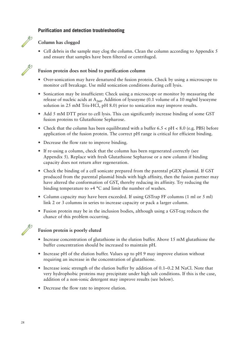
28 Purification and detection troubleshooting Column has clogged • Cell debris in the sample may clog the column. Clean the column according to Appendix 5 and ensure that samples have been filtered or centrifuged. Fusion protein does not bind to purification column • Over-sonication may have denatured the fusion protein. Check by using a microscope to monitor cell breakage. Use mild sonication conditions during cell lysis. • Sonication may be insufficient: Check using a microscope or monitor by measuring the release of nucleic acids at A260. Addition of lysozyme (0.1 volume of a 10 mg/ml lysozyme solution in 25 mM Tris-HCl, pH 8.0) prior to sonication may improve results. • Add 5 mM DTT prior to cell lysis. This can significantly increase binding of some GST fusion proteins to Glutathione Sepharose. • Check that the column has been equilibrated with a buffer 6.5 < pH < 8.0 (e.g. PBS) before application of the fusion protein. The correct pH range is critical for efficient binding. • Decrease the flow rate to improve binding. • If re-using a column, check that the column has been regenerated correctly (see Appendix 5). Replace with fresh Glutathione Sepharose or a new column if binding capacity does not return after regeneration. • Check the binding of a cell sonicate prepared from the parental pGEX plasmid. If GST produced from the parental plasmid binds with high affinity, then the fusion partner may have altered the conformation of GST, thereby reducing its affinity. Try reducing the binding temperature to +4 °C and limit the number of washes. • Column capacity may have been exceeded. If using GSTrap FF columns (1 ml or 5 ml) link 2 or 3 columns in series to increase capacity or pack a larger column. • Fusion protein may be in the inclusion bodies, although using a GST-tag reduces the chance of this problem occurring. Fusion protein is poorly eluted • Increase concentration of glutathione in the elution buffer. Above 15 mM glutathione the buffer concentration should be increased to maintain pH. • Increase pH of the elution buffer. Values up to pH 9 may improve elution without requiring an increase in the concentration of glutathione. • Increase ionic strength of the elution buffer by addition of 0.1–0.2 M NaCl. Note that very hydrophobic proteins may precipitate under high salt conditions. If this is the case, addition of a non-ionic detergent may improve results (see below). • Decrease the flow rate to improve elution

÷Adao咖derergent01%m0oor2光ha my rev2nc,c c hydrophobic c interactions .Try over-night elution at room temperature. Multiple bands seen on SDS-PAGE or Western blot analysis and co-purification ost prote wth the by sion protein .Check that a protease-deficient host such as E.coli B21 has been used. .Add protease inhibitors such as 1 mM PMSF to the lysis solution.A non-toxic,water soluble alternative to PMSF is 4-(2-amino-ethyl)-benzenesulfonyl fluoride hydrochloride (AEBSF),commercially available as PefablocT SC from Boehringer Mannheim. Use prepacked GSTrap FF columns or Glutathione Sepharose 4 Fast Flow.These can be used at higher flow rates to process samples more quickly and so avoid degradation. Decrease sonication.Addition of lysozyme (0.1 volume of a 10 mg/ml lysozyme solution in 25 mM Tris-HCl,pH 8.0)prior to sonication may improve results.Avoid frothing as this may denature the fusion protein. step (s Chapter )A variety of proreins known as g or nascent proteins I coli may co-purify with GST fusion proteins,includinga M,000 protein (see below). Serine protease inhibitors must be removed prior to cleavage by thrombin or Factor Xa. Use HiTrap Benzamidine FF(high sub)(see page 39). M,70 000 protein co-purifies with the GST-fusion protein Pre incubate the protein solution with 2 mM ATP,10 mM mM Tris-HCI (pH 7.4)for 10 tion in order dis the mplex This M.70 000 is probably a p of the E.oli gen and inv olved in the d of "al eins in E.coli.Re be ren P uggest nat this tein can ns p24.1 A sham Pha metbods.199406/0813.btmll)or by (Myers,M.,BIOSCI posting 71l1q93 Thain,A.,et al.Trends Genet.12,209-210 (1996)and Sherman,M.and Goldberg.A.L. 1.Biol.Che .269.31479-31483,(1994)suggest washing the column with ATP or GroES rather than using a subsequent IEX step 29
29 • Add a non-ionic detergent (0.1% Triton™ X-100 or 2% N-octyl glucoside) to the elution buffer to reduce non-specific hydrophobic interactions that may prevent solubilization and elution of fusion proteins • Try over-night elution at room temperature or +4 °C. Multiple bands seen on SDS-PAGE or Western blot analysis Multiple bands result from partial degradation of fusion proteins by proteases, or denaturation and co-purification of host proteins with the GST fusion protein due to over-sonication. • Check that a protease-deficient host such as E. coli B21 has been used. • Add protease inhibitors such as 1 mM PMSF to the lysis solution. A non-toxic, water soluble alternative to PMSF is 4-(2-amino-ethyl)- benzenesulfonyl fluoride hydrochloride (AEBSF), commercially available as Pefabloc™ SC from Boehringer Mannheim. • Use prepacked GSTrap FF columns or Glutathione Sepharose 4 Fast Flow. These can be used at higher flow rates to process samples more quickly and so avoid degradation. • Decrease sonication. Addition of lysozyme (0.1 volume of a 10 mg/ml lysozyme solution in 25 mM Tris-HCl, pH 8.0) prior to sonication may improve results. Avoid frothing as this may denature the fusion protein. • Include an additional purification step (see Chapter 9). A variety of proteins known as chaperonins that are involved in the correct folding of nascent proteins in E. coli may co-purify with GST fusion proteins, including a Mr 70 000 protein (see below). Serine protease inhibitors must be removed prior to cleavage by thrombin or Factor Xa. Use HiTrap Benzamidine FF (high sub) (see page 39). Mr 70 000 protein co-purifies with the GST-fusion protein Pre-incubate the protein solution with 2 mM ATP, 10 mM MgSO4, 50 mM Tris-HCl (pH 7.4) for 10 minutes at +37 °C prior to purification in order to dissociate the complex. This Mr 70 000 protein is probably a protein product of the E. coli gene dnaK and involved in the degradation of "abnormal" proteins in E. coli. Reports suggest that this protein can be removed by ion exchange chromatography (Analects and Separations p24, 1996, Amersham Pharmacia Biotech and (http://bionet.hgmp.mrc.ac.uk/hypermail/methods/ methods.199406/0813.html)) or by passage of the sample over ATP agarose (Myers, M., BIOSCI posting, 7 July 1993). Thain, A., et al. Trends Genet. 12, 209–210 (1996) and Sherman, M. and Goldberg, A.L., J. Biol. Chem, 269, 31479–31483, (1994) suggest washing the column with ATP or GroES rather than using a subsequent IEX step

Tag removal by enzymatic cleavage In most cases functional tests can be performed using the intact fusion with gst if removal of the GST tag is necessary,it is highly re ommended to pr roduce the fusion proteins with site.The GST tag then can be re in a single step on the column (sce Figure 7).This protease also has the useful rty of active at +4C thus allowing cle age to be performed at low emperature Thrombin or Factor Xa recognition sites may be cleaved either while bound on the column or in solution after elution from the column (see figure 8).The protease used for cleavage can be removed using Benzamidine Sepharose,a purification medium with a high specificity for serine proteases (see page 39). On-column cleavage is generally recommended as the method of choice since ineluted a HiTrap Benzamidine FF (high sub) lumn can be nected in se ies so that cle ed he gSTr ap FE the HiTrap Benza idine FF(high sub) The amount of enzyme,temperature and length of incubation required for complete digestion varies according to the specific GST fusion protein produced.Determine optimal conditions in pilot experiments. Remove samples at various time points and analyse by SDS-PAGE to estimate the yield, purity and extent of digestion.Approximate molecular weights for SDS-PAGE analysis PreScission Protease M37000 Bovine Factor Xa M,48000 If protease inhibitors have been used in the lysis soluti on the Xa (the ey must be rer r to e by PreSciss th oedprior Iohihitor Factor Xa and thrombin Cleavage of fusion proteins is most commonly performed on milligram quantities of fusion protein suitable for purification on GSTrap FE The following protocols describe a manual cleavage and purification using a syringe and a 1 ml GSTrap FF column.The protocols can be adapted for use with GST MicroSpin columns to work at smaller scales or scaled up onto larger columns to run on AKTAdesign systems
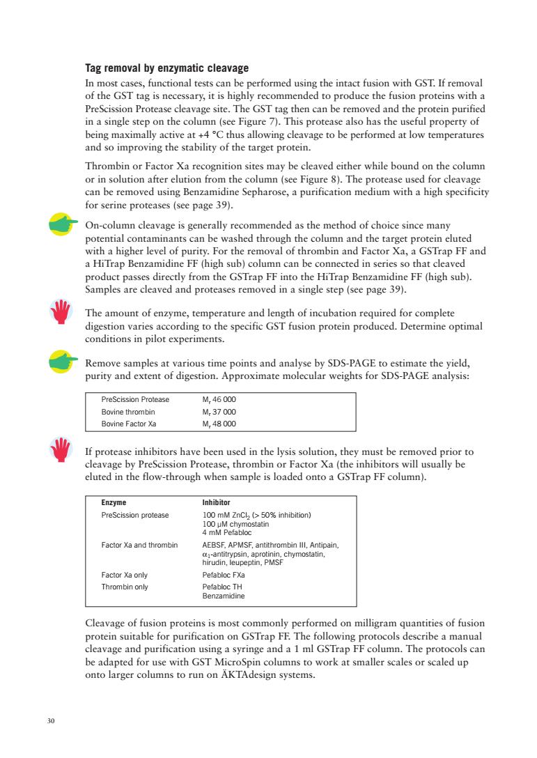
30 PreScission Protease Mr 46 000 Bovine thrombin Mr 37 000 Bovine Factor Xa Mr 48 000 Tag removal by enzymatic cleavage In most cases, functional tests can be performed using the intact fusion with GST. If removal of the GST tag is necessary, it is highly recommended to produce the fusion proteins with a PreScission Protease cleavage site. The GST tag then can be removed and the protein purified in a single step on the column (see Figure 7). This protease also has the useful property of being maximally active at +4 °C thus allowing cleavage to be performed at low temperatures and so improving the stability of the target protein. Thrombin or Factor Xa recognition sites may be cleaved either while bound on the column or in solution after elution from the column (see Figure 8). The protease used for cleavage can be removed using Benzamidine Sepharose, a purification medium with a high specificity for serine proteases (see page 39). On-column cleavage is generally recommended as the method of choice since many potential contaminants can be washed through the column and the target protein eluted with a higher level of purity. For the removal of thrombin and Factor Xa, a GSTrap FF and a HiTrap Benzamidine FF (high sub) column can be connected in series so that cleaved product passes directly from the GSTrap FF into the HiTrap Benzamidine FF (high sub). Samples are cleaved and proteases removed in a single step (see page 39). The amount of enzyme, temperature and length of incubation required for complete digestion varies according to the specific GST fusion protein produced. Determine optimal conditions in pilot experiments. Remove samples at various time points and analyse by SDS-PAGE to estimate the yield, purity and extent of digestion. Approximate molecular weights for SDS-PAGE analysis: If protease inhibitors have been used in the lysis solution, they must be removed prior to cleavage by PreScission Protease, thrombin or Factor Xa (the inhibitors will usually be eluted in the flow-through when sample is loaded onto a GSTrap FF column). Enzyme Inhibitor PreScission protease 100 mM ZnCl2 (> 50% inhibition) 100 µM chymostatin 4 mM Pefabloc Factor Xa and thrombin AEBSF, APMSF, antithrombin III, Antipain, a1-antitrypsin, aprotinin, chymostatin, hirudin, leupeptin, PMSF Factor Xa only Pefabloc FXa Thrombin only Pefabloc TH Benzamidine Cleavage of fusion proteins is most commonly performed on milligram quantities of fusion protein suitable for purification on GSTrap FF. The following protocols describe a manual cleavage and purification using a syringe and a 1 ml GSTrap FF column. The protocols can be adapted for use with GST MicroSpin columns to work at smaller scales or scaled up onto larger columns to run on ÄKTAdesign systems