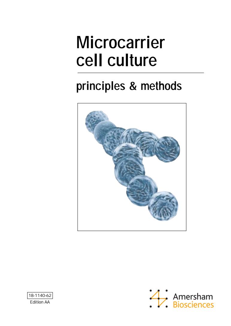
Microcarrier cell culture principles methods 18-1140-62 找 Amersham Edition AA Biosciences
Microcarrier cell culture principles & methods 18-1140-62 Edition AA
Microcarrier cell culture principles methods
Microcarrier cell culture principles & methods
Contents 1.Background. 1.1 Introduction 7 1.2.Adhesion of cells to culture surfaces 1.3.The development of microcarriers for animal cell culture 11 1.4.Applications of microcarrier culture. 1.4.1.Cell types cultured on Cytodex microcarriers. 15 1.4.2.Production of large numbers of cells. 7 1.4.3.Production of viruses and cell products 18 1.4.4.Studies on cell function,metabolism and differentiation. .22 1.4.5.Proteolytic enzyme-free subcultivation and cell transfer.25 1.4.6.Microscopy. .26 1.4.7.Harvesting mitotic cells. .26 1.4.8.Transportation and storage of cells. .27 2.Cytodex microcarriers. . 2.1.Requirements for an optimum microcarrier 29 2.2.Cytodex1 30 2.3.Cytodex 3. .30 2结:wth9 dor1otc7 34 .35 3.Microcarrier cell culture methods 3.1.General outline of procedure .3 3.2.Microcarrier culture vessels 3.2.1.Requirements. 3.2.2 aboratory scale microcarrier culture vessels. 3.2.3 rge scale microcarrier culture vessels 3.2.4.Siliconizing culture vessels Initial stirring. 3.4.3 Concentration of microcarriers. gmoahipbetcapatgemtagyaiaciaepocoahnk during the initial culture ph 3.5 aining a mic crocarrier culture . 352 88 tof cultu 3.53 re medium Intaining cultures at confluence
Contents 1. Background . 7 1.1 Introduction . 7 1.2 . Adhesion of cells to culture surfaces . 9 1.3 . The development of microcarriers for animal cell culture. 11 1.4 . Applications of microcarrier culture. 14 1.4.1 . Cell types cultured on Cytodex microcarriers . 15 1.4.2 . Production of large numbers of cells . 17 1.4.3 . Production of viruses and cell products . 18 1.4.4 . Studies on cell function, metabolism and differentiation . 22 1.4.5 . Proteolytic enzyme-free subcultivation and cell transfer . 25 1.4.6 . Microscopy. 26 1.4.7 . Harvesting mitotic cells . 26 1.4.8 . Transportation and storage of cells . 27 2. Cytodex microcarriers . 29 2.1 . Requirements for an optimum microcarrier . 29 2.2 . Cytodex 1 . 30 2.3 . Cytodex 3 . 30 2.4 . Which Cytodex microcarrier to use? . 34 2.5 . Availability and storage . 35 3. Microcarrier cell culture methods . 37 3.1 . General outline of procedure . 37 3.2 . Microcarrier culture vessels . 38 3.2.1 . Requirements . 38 3.2.2 . Laboratory scale microcarrier culture vessels. 39 3.2.3 . Large scale microcarrier culture vessels . 45 3.2.4 . Siliconizing culture vessels . 45 3.3 . Preparing Cytodex microcarriers for culture . 46 3.4 . Initiating a microcarrier culture . 47 3.4.1 . Equilibration before inoculation . 47 3.4.2 . Initial stirring. 48 3.4.3 . Concentration of microcarriers . 51 3.4.4 . Inoculation density . 52 3.4.5 . Inoculum condition. 53 3.4.6 . Culture media during the initial culture phase . 54 3.4.7 . Relationship between plating efficiency and culture procedure56 3.5 . Maintaining a microcarrier culture . 58 3.5.1 . Stirring speed . 58 3.5.2 . Replenishment of culture medium . 61 3.5.3 . Maintaining cultures at confluence . 65
3.6 Monitoring the growth of cells and microscopy 36 1 Dir unting cells relea opy 44444 3 63 Countin released nuclei 36 4 Fivin 87 3.6.5.Staining cells cells and subculturing 78 3.7.4.Cold treatment. 81 3.7.5.Sonication. 8I 3.7.6.Lignocaine for harvesting macrophages .7.7.Modifications to harvesting procedures forlarge scale cres 378c parating detached cells fro 3.7.9.Measurement of cell viability 82 3.7.10.Subculturing techniques 83 3 7 11 Re-use of Cvtodex 84 4.General considerations. 86 4.1.Culture media .85 4.1.1.Choice of culture medium 85 4.1.2.General comments on components of culture media 88 4.1.3.Practical aspects of culture media .91 4.2.Serum supplements .91 4.2.1.The purpose of serum in culture media .91 4.2.2.Choice and concentration of serum supplement. 92 4.2.3.Variability of sera .96 4.2.4.Serum free media .97 4.3.Gas supply. .97 4.3.1 Gas supply and exchange in microcarrier cultures. .98 4.3.2.Oxygen .99 4.3.3.Carbon dioxide. .99 4.3.4 Purity of the gas supply 100 4.4 Culture pH. .100 4.4.1.pH optima for cell culture 100 4.4.2.Buffers and the control of pH. 10 4.4.3.Minimizing accumulation of lactate 104 4.5.Osmolarity. 105 4.6 Frezing cells for storage 106 4.6.1 Procedure for freezing and thawing 106 4.6.2 Storage medium. 10 4.7 Contamination . 107
3.6 . Monitoring the growth of cells and microscopy . 66 3.6.1 . Direct observation by microscopy. 66 3.6.2 . Counting cells released after trypsinization . 67 3.6.3 . Counting released nuclei . 67 3.6.4 . Fixing cells. 67 3.6.5 . Staining cells . 76 3.7 . Harvesting cells and subculturing . 77 3.7.1 . Chelating agents . 78 3.7.2 . Proteolytic enzymes . 78 3.7.3 . Hypotonic treatment. 80 3.7.4 . Cold treatment. 81 3.7.5 . Sonication. 81 3.7.6 . Lignocaine for harvesting macrophages . 81 3.7.7 . Modifications to harvesting procedures for large scale cultures 81 3.7.8 . Separating detached cells from microcarriers . 82 3.7.9 . Measurement of cell viability . 82 3.7.10 . Subculturing techniques . 83 3.7.11 . Re-use of Cytodex . 84 4. General considerations. 85 4.1 . Culture media . 85 4.1.1 . Choice of culture medium . 85 4.1.2 . General comments on components of culture media . 88 4.1.3 . Practical aspects of culture media . 91 4.2 . Serum supplements . 91 4.2.1 . The purpose of serum in culture media . 91 4.2.2 . Choice and concentration of serum supplement . 92 4.2.3 . Variability of sera . 96 4.2.4 . Serum free media . 97 4.3 . Gas supply . 97 4.3.1 Gas supply and exchange in microcarrier cultures . 98 4.3.2 . Oxygen . 99 4.3.3 . Carbon dioxide. 99 4.3.4 Purity of the gas supply . 100 4.4 Culture pH. 100 4.4.1 . pH optima for cell culture. 100 4.4.2 . Buffers and the control of pH . 102 4.4.3 . Minimizing accumulation of lactate . 104 4.5 . Osmolarity. 105 4.6 Frezing cells for storage . 106 4.6.1 Procedure for freezing and thawing . 106 4.6.2 Storage medium . 107 4.7 Contamination . 107
5.Optimizing culture conditions and trouble shooting .111 6.Appendix. 115 6.1 Cells cultured on Cytodex microcarriers .115 6.2 Examples of microcarrier culture protocols .121 6.2.1 Diploid human fibroblast and the production of interferon.121 6.2.2 African Green monkey kidney cells (Vero)and the production of Simian Virus 40. 。 122 6.2.3 Primary monkey or dog kidney cells 123 6.2.4 Primary chicken embryo fibroblasts 12 6.2.5 Baby hamster kidney cells (BHK) .125 6.3 Methods for determining the protein and DNA content of cells grown on microcarriers. 6.4 Abbreviations .126 7.References 127
5. Optimizing culture conditions and trouble shooting . 111 6. Appendix . 115 6.1 Cells cultured on Cytodex microcarriers . 115 6.2 Examples of microcarrier culture protocols. 121 6.2.1 Diploid human fibroblast and the production of interferon . 121 6.2.2 African Green monkey kidney cells (Vero) and the production of Simian Virus 40 . 122 6.2.3 Primary monkey or dog kidney cells . 123 6.2.4 Primary chicken embryo fibroblasts . 123 6.2.5 Baby hamster kidney cells (BHK) . 125 6.3 Methods for determining the protein and DNA content of cells grown on microcarriers. 125 6.4 Abbreviations . 126 7. References . 127