Chapter II Pyrrophyta(甲藻门) Department of Oceanography Xiamen University Mesokaryotes: nucleus enveloped the nuclear membrane (Eukaryotes), but has no histone and its chromatin is circular (Monera)
Chapter II Pyrrophyta(甲藻门) Department of Oceanography Xiamen University Mesokaryotes: nucleus enveloped the nuclear membrane (Eukaryotes), but has no histone and its chromatin is circular (Monera)
References • Hallegraeff G. M. et al., 1995. Manual on harmful marine microalgae. UNESCO. 551p • Tomas C. R., 1997. Identifying marine phytoplankton. Academic Press. 858p • Garces E. et al., 2001. Life histories of microalgal species causing harmful blooms. 208p • Botes L., 2003. Phytoplankton identification catalogue – Saldanha Bay, South Africa, April 2001. GloBallast Monograph Series, 7. IMO London. 87p • 国家海洋环境监测中心 《中国近海赤潮生物图谱——简 本》
References • Hallegraeff G. M. et al., 1995. Manual on harmful marine microalgae. UNESCO. 551p • Tomas C. R., 1997. Identifying marine phytoplankton. Academic Press. 858p • Garces E. et al., 2001. Life histories of microalgal species causing harmful blooms. 208p • Botes L., 2003. Phytoplankton identification catalogue – Saldanha Bay, South Africa, April 2001. GloBallast Monograph Series, 7. IMO London. 87p • 国家海洋环境监测中心 《中国近海赤潮生物图谱——简 本》
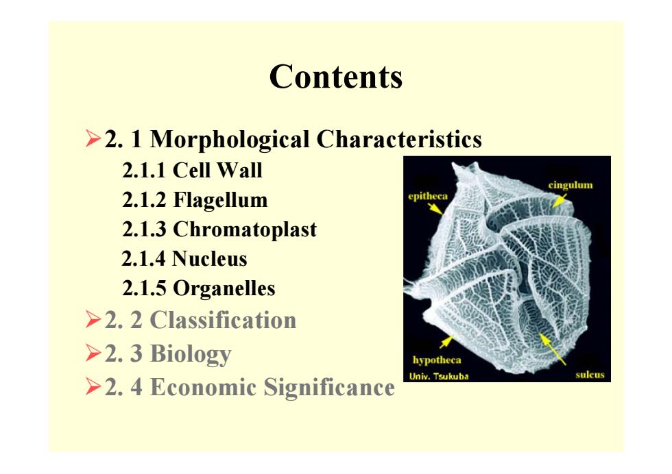
Contents ¾2. 1 Morphological Characteristics 2.1.1 Cell Wall 2.1.2 Flagellum 2.1.3 Chromatoplast 2.1.4 Nucleus 2.1.5 Organelles ¾2. 2 Classification ¾2. 3 Biology ¾2. 4 Economic Significance
Contents ¾2. 1 Morphological Characteristics 2.1.1 Cell Wall 2.1.2 Flagellum 2.1.3 Chromatoplast 2.1.4 Nucleus 2.1.5 Organelles ¾2. 2 Classification ¾2. 3 Biology ¾2. 4 Economic Significance
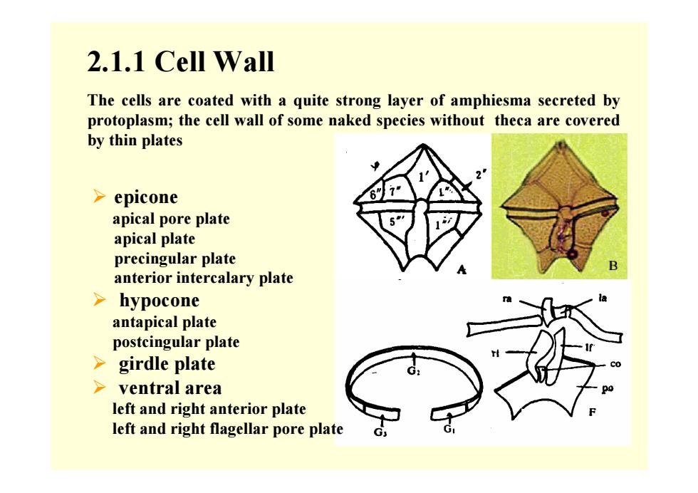
2.1.1 Cell Wall ¾ epicone apical pore plate apical plate precingular plate anterior intercalary plate ¾ hypocone antapical plate postcingular plate ¾ girdle plate ¾ ventral area left and right anterior plate left and right flagellar pore plate The cells are coated with a quite strong layer of amphiesma secreted by protoplasm; the cell wall of some naked species without theca are covered by thin plates
2.1.1 Cell Wall ¾ epicone apical pore plate apical plate precingular plate anterior intercalary plate ¾ hypocone antapical plate postcingular plate ¾ girdle plate ¾ ventral area left and right anterior plate left and right flagellar pore plate The cells are coated with a quite strong layer of amphiesma secreted by protoplasm; the cell wall of some naked species without theca are covered by thin plates
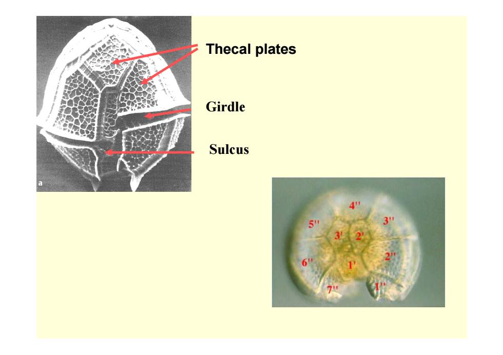
Thecal plates Girdle Sulcus
Thecal plates Girdle Sulcus