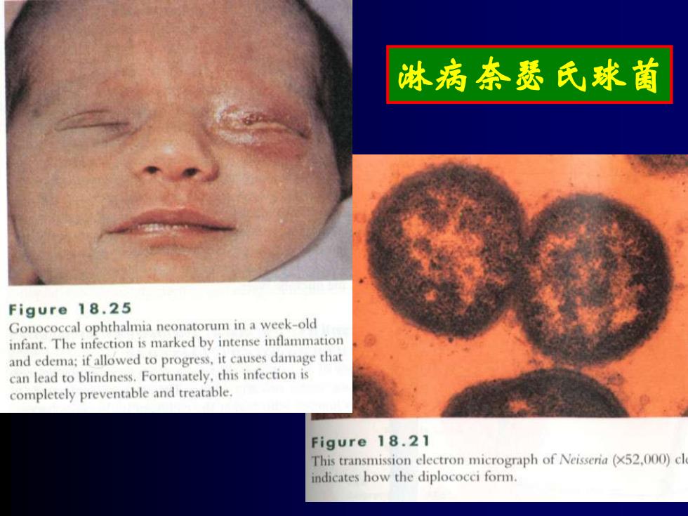
淋病奈瑟氏球菌 Figure 18.25 Gonococcal ophthalmia neonatorum in a week-old infant.The infection is marked by intense inflammation and edema;if allowed to progress,it causes damage that can lead to blindness.Fortunately,this infection is completely preventable and treatable. Figure 18.21 This transmission electron micrograph of Neisseria(x52,000)cl indicates how the diplococci form
淋病奈瑟氏球菌
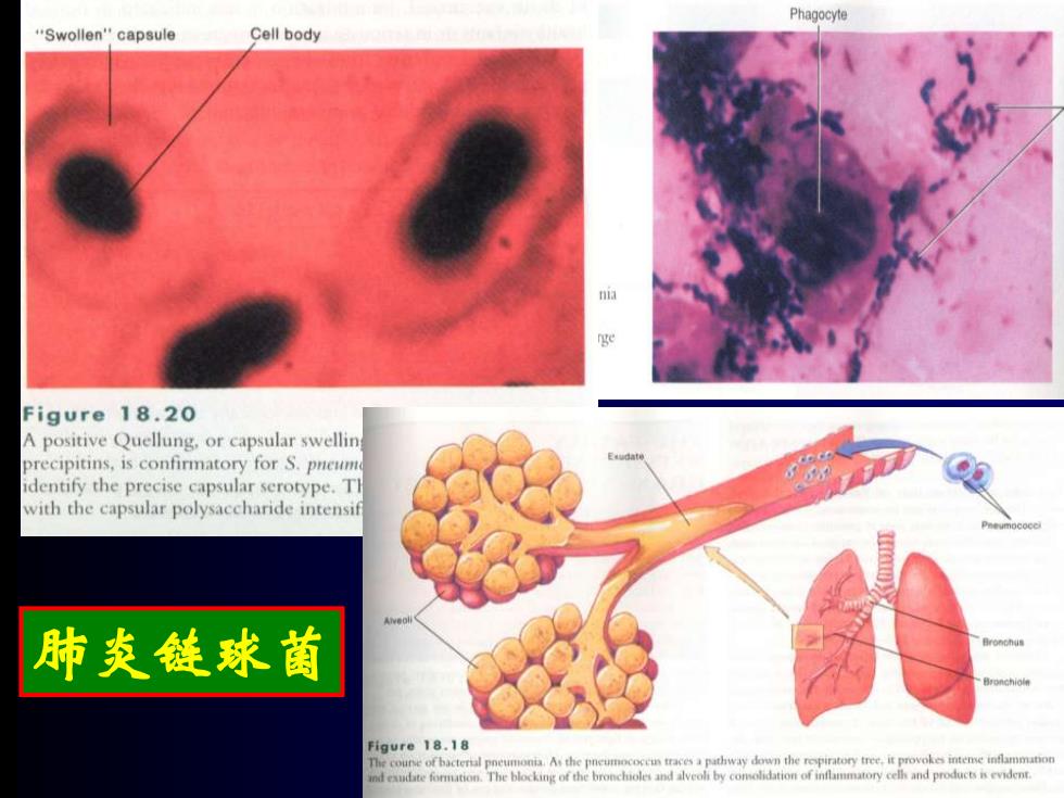
Phagocyte "Swollen"capsule Cell body a ge Figure 18.20 A positive Quellung,or capsular swellin precipitins,is confirmatory for S.pneum identify the precise capsular serotype.Tl with the capsular polysaccharide intensif 肺炎链球菌 Figure 18.18 The cou of bacteral A the pathway down the repiratory tree.it provokes inteme ind eaudte formation.The blocking of the broncholes and alveoh by comolidation of inflammatory celh and products is evident
肺炎链球菌
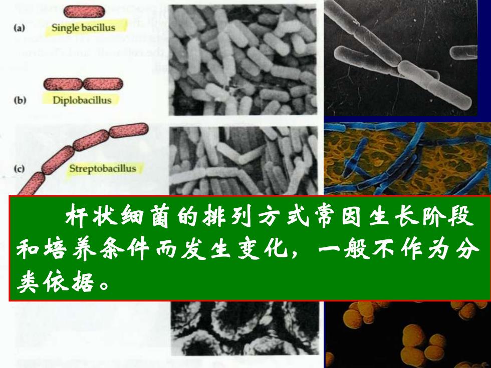
(a) Single bacillus (b) Diplobacillus (c) Streptobacillus 杆状细菌的排列方式常因生长阶段 和培养条件而发生变化,一殷不作为分 类依据
一、一般形态及细胞结构 (一)个体形态和排列 2、杆状 细胞呈杆状或圆柱形,一般其粗细(直 径)比较稳定,而长度则常因培养时间、培 养条件不同而有较大变化。 杆状细菌的排列方式常因生长阶段 和培养条件而发生变化,一般不作为分 类依据
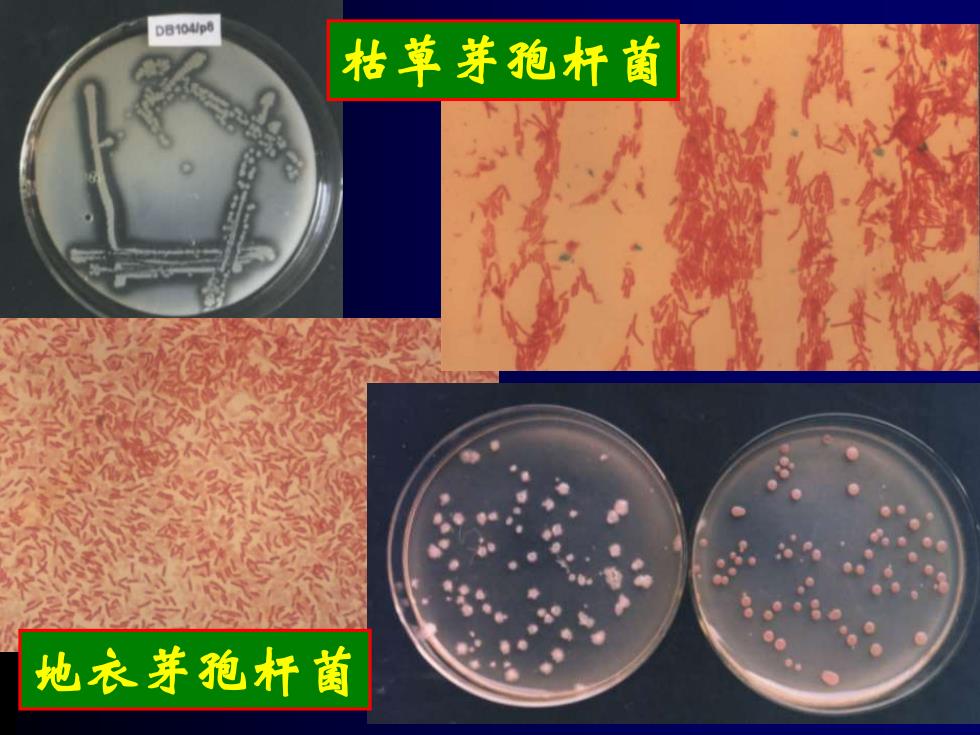
DB104/p 枯草芽孢杆菌 地衣芽孢杆菌
枯草芽孢杆菌 地衣芽孢杆菌
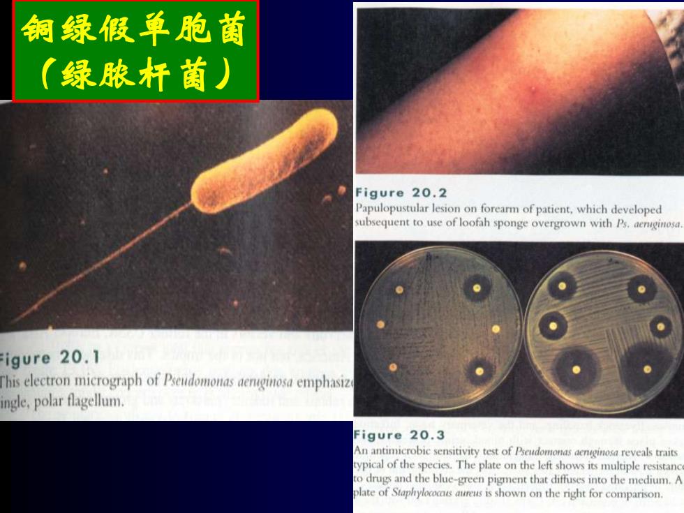
铜绿假单胞菌 (绿脓杆菌) Figure 20.2 Papulopustular lesion on forearm of patient,which developed subsequent to use of loofah sponge overgrown with Ps.aenginosa igure 20.1 This electron micrograph of Pudomonsn emphasiz ingle,polar flagellum. Figure 20.3 An antimicrobic sensitivity test of Pseudomonas aenginosa reveals traits ypical of the species.The plate on the left shows its multiple resistanc to drugs and the blue-green pigment that diffuses into the medium.A olate of Staphyloccs aureus is shown on the right for comparison
铜绿假单胞菌 (绿脓杆菌)