Glass spheres were not suitable because their high density required stirring speeds for suspension which were not compatible with cell growth(17.31).Several workers have suggested that the ideal microcarrier should have properties similar to those of DEAE-Sephadex A-50(16.22.32-35).Other ion-exchange beads all proved to be inferior to DEAE-Sephadex A-50(13,14,16). Using DEAE-Sephadex A-50 at a concentration of 1 mg/ml van Wezel demonstrated that a homogeneous microcarriersystem could be used for the large scale culture of anchorage-dependent cells,including diploid human fibroblasts(31).This early work illustrated the potential of the microcarrier technique for producing virus and latter experiments established that this technique could be scaled-up for a variety of large-scale production processes (1,33). Since the yield of anchorage-dependent cells depends on the surface area available for growth.it was believed that the maximum cell density (yield)in microcarrier cultures wo depend on the microcarrier surface area(1).However,when the atoipEdA0m2a82issedd A-50 exceedec The explanatio an to rge a tota city in Substitution(meq/g) g The relaionshp etheo ublished work from 12
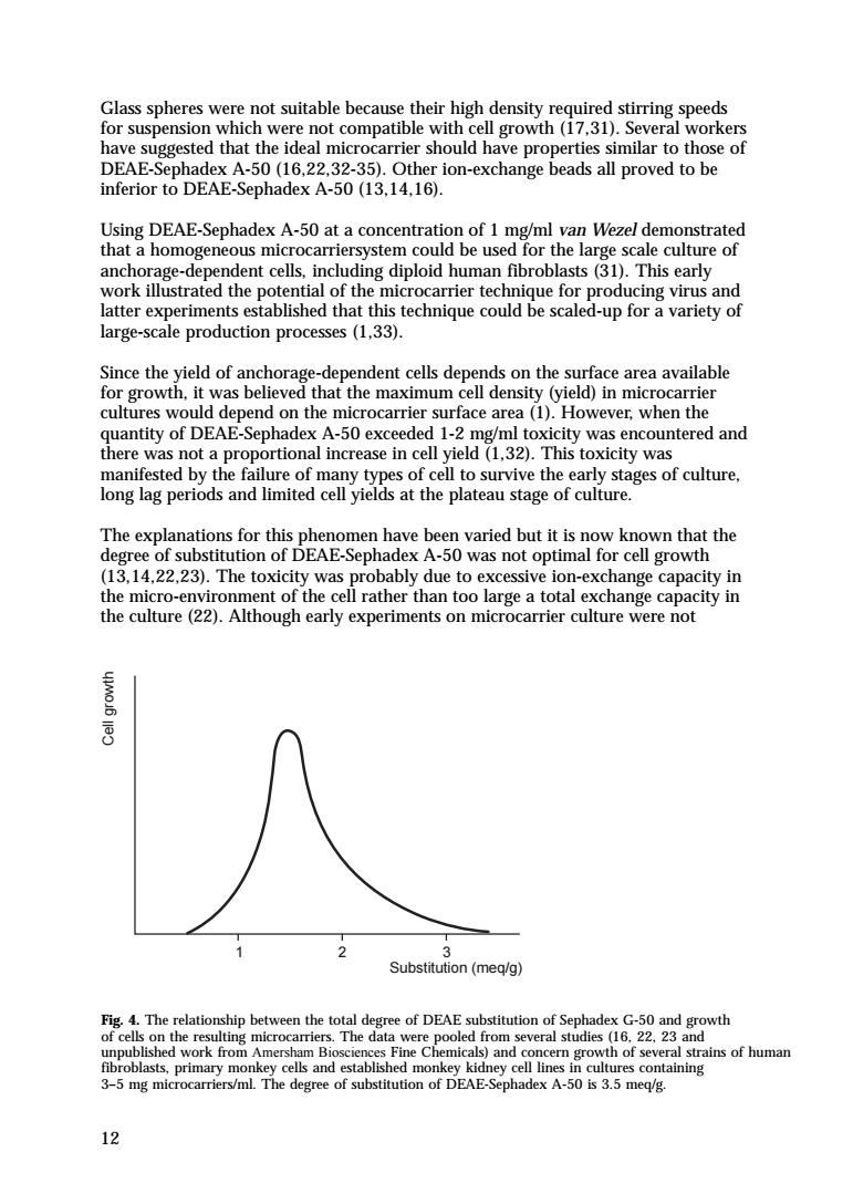
12 Glass spheres were not suitable because their high density required stirring speeds for suspension which were not compatible with cell growth (17,31). Several workers have suggested that the ideal microcarrier should have properties similar to those of DEAE-Sephadex A-50 (16,22,32-35). Other ion-exchange beads all proved to be inferior to DEAE-Sephadex A-50 (13,14,16). Using DEAE-Sephadex A-50 at a concentration of 1 mg/ml van Wezel demonstrated that a homogeneous microcarriersystem could be used for the large scale culture of anchorage-dependent cells, including diploid human fibroblasts (31). This early work illustrated the potential of the microcarrier technique for producing virus and latter experiments established that this technique could be scaled-up for a variety of large-scale production processes (1,33). Since the yield of anchorage-dependent cells depends on the surface area available for growth, it was believed that the maximum cell density (yield) in microcarrier cultures would depend on the microcarrier surface area (1). However, when the quantity of DEAE-Sephadex A-50 exceeded 1-2 mg/ml toxicity was encountered and there was not a proportional increase in cell yield (1,32). This toxicity was manifested by the failure of many types of cell to survive the early stages of culture, long lag periods and limited cell yields at the plateau stage of culture. The explanations for this phenomen have been varied but it is now known that the degree of substitution of DEAE-Sephadex A-50 was not optimal for cell growth (13,14,22,23). The toxicity was probably due to excessive ion-exchange capacity in the micro-environment of the cell rather than too large a total exchange capacity in the culture (22). Although early experiments on microcarrier culture were not Cell growth Substitution (meq/g) 1 2 3 Fig. 4. The relationship between the total degree of DEAE substitution of Sephadex G-50 and growth of cells on the resulting microcarriers. The data were pooled from several studies (16, 22, 23 and unpublished work from Amersham Biosciences Fine Chemicals) and concern growth of several strains of human fibroblasts, primary monkey cells and established monkey kidney cell lines in cultures containing 3–5 mg microcarriers/ml. The degree of substitution of DEAE-Sephadex A-50 is 3.5 meq/g
16.0 Cytodex 影h Key K Monolayer 1.0 025 100 200 300 Hours controlled or optimized for various culture parameters such as plating efficiency. inoculation density.serum and culture medium,the work of Kuchler et al(36). Inooka(37)and Horng and McLimans(13,14)suggested that alterations in the ion-exchange capacity of DEAE-Sephadex A-50 could lead to improvements in cell attachment and growth.The ion-exchange capacity of DEAE-Sephadex A-50 could be altered by changing the culture environment (e.g.ionic strength.pH)but such changes were very limited since cells require physiological conditions for growth. This problem was overcome by the development of microcarriers with a much lower degree of substitution than DEAE-Sephadex A-50 and which also fulfilled the requirements for an optimal microcarrier(16,22,23,33,35). Figure 4 shows the effect that different degrees of substitution of Sephadex with DEAE-chloride has on cell growth.Using Sephadex as the starting material and substituting the matrix with DEAE groups to 1.5 meq/g dry product.it was possible to achieve a microcarrier,Cytodex 1.which was suitable for the growth of a wide ciated with E-Sephadex A-50 are avoided and microcarrier concentrations well in excess of 1 mg/ml can be used with concomitant increases in cell yield. Hence this reduced charge microcarrier was the first product to allow the full timal microcarrier (16.17,section s are used th f most cells on Cvtodex microcarrier is comparable to that achieved on pastic or gass cul surfaces (fig.5). 13
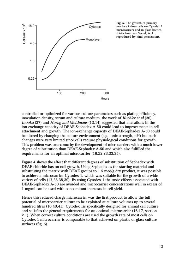
13 controlled or optimized for various culture parameters such as plating efficiency, inoculation density, serum and culture medium, the work of Kuchler et al (36), Inooka (37) and Horng and McLimans (13,14) suggested that alterations in the ion-exchange capacity of DEAE-Sephadex A-50 could lead to improvements in cell attachment and growth. The ion-exchange capacity of DEAE-Sephadex A-50 could be altered by changing the culture environment (e.g. ionic strength, pH) but such changes were very limited since cells require physiological conditions for growth. This problem was overcome by the development of microcarriers with a much lower degree of substitution than DEAE-Sephadex A-50 and which also fulfilled the requirements for an optimal microcarrier (16,22,23,33,35). Figure 4 shows the effect that different degrees of substitution of Sephadex with DEAE-chloride has on cell growth. Using Sephadex as the starting material and substituting the matrix with DEAE groups to 1.5 meq/g dry product, it was possible to achieve a microcarrier, Cytodex 1, which was suitable for the growth of a wide variety of cells (17,23,38,39). By using Cytodex 1 the toxic effects associated with DEAE-Sephadex A-50 are avoided and microcarrier concentrations well in excess of 1 mg/ml can be used with concomitant increases in cell yield. Hence this reduced charge microcarrier was the first product to allow the full potential of microcarrier culture to be exploited at culture volumes up to several hundred litres (10,40,41). Cytodex 1is specifically designed for animal cell culture and satisfies the general requirements for an optimal microcarrier (16,17, section 2.1). When correct culture conditions are used the growth rate of most cells on Cytodex 1 microcarrier is comparable to that achieved on plastic or glass culture surfaces (fig. 5). 100 200 300 Hours 0.25 1.0 4.0 16.0 Cells/ml x 105 Monolayer Cytodex Fig. 5. The growth of primary monkey kidney cells on Cytodex 1 microcarriers and in glass bottles. (Data from van Wezel, A. L., reprodused by kind permission)
The development of Cytodex 1 microcarriers has taken into account the requirements for attachment of cells(section 1.2)and the procedures necessary for maximum growth of a wide variety of cells in microcarrier culture(section 3).The possibilities for microcarrier culture of animal cells have been increased further by the development of Cytodex 2 and Cytodex 3 microcarriers(section 2.3 and 2.4). Since charged groups are necessary only for cell attachment they need only be confined to the surface of the microcarriers. Cytodex 3 microcarriers represent a new concept in microcarrier culture.Instead of amechog2nTme6egwahk8ae using syntethic charged groups to promote cell attachment,thes hrgeadghtcacicanei5pgmamaseYog d aemmpe ulur The relative properties and uses of the various types of Cytodex microcarriers are outlined in section 2. 1.4 Applications of microcarrier culture nd the e app cate b) tudie uts in s on cotic vitro an cell ulture a very wi ariety nt c ap re ddit ation can be obtaine d from ersham Fig.6.Human-mouse hybrid growing o parental s and HGRT humar by kind permiso 14
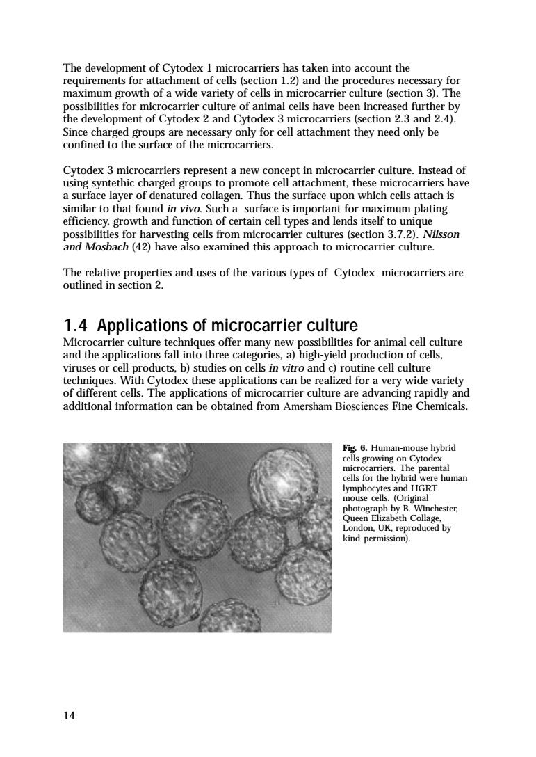
14 The development of Cytodex 1 microcarriers has taken into account the requirements for attachment of cells (section 1.2) and the procedures necessary for maximum growth of a wide variety of cells in microcarrier culture (section 3). The possibilities for microcarrier culture of animal cells have been increased further by the development of Cytodex 2 and Cytodex 3 microcarriers (section 2.3 and 2.4). Since charged groups are necessary only for cell attachment they need only be confined to the surface of the microcarriers. Cytodex 3 microcarriers represent a new concept in microcarrier culture. Instead of using syntethic charged groups to promote cell attachment, these microcarriers have a surface layer of denatured collagen. Thus the surface upon which cells attach is similar to that found in vivo. Such a surface is important for maximum plating efficiency, growth and function of certain cell types and lends itself to unique possibilities for harvesting cells from microcarrier cultures (section 3.7.2). Nilsson and Mosbach (42) have also examined this approach to microcarrier culture. The relative properties and uses of the various types of Cytodex microcarriers are outlined in section 2. 1.4 Applications of microcarrier culture Microcarrier culture techniques offer many new possibilities for animal cell culture and the applications fall into three categories, a) high-yield production of cells, viruses or cell products, b) studies on cells in vitro and c) routine cell culture techniques. With Cytodex these applications can be realized for a very wide variety of different cells. The applications of microcarrier culture are advancing rapidly and additional information can be obtained from Amersham Biosciences Fine Chemicals. Fig. 6. Human-mouse hybrid cells growing on Cytodex microcarriers. The parental cells for the hybrid were human lymphocytes and HGRT mouse cells. (Original photograph by B. Winchester, Queen Elizabeth Collage, London, UK, reproduced by kind permission)
e spleen and human on Cytodex umour Too Lond Ho by kind permission) d9a 1.4.1 Cell types cultured on Cytodex microcarriers The cell types successfully cultured on microcarriers are listed in Section 6.1.and examples of virtually all classes of cultured animal cells are represented.Cytodex microcarriers are cell culture surfaces of general applicability and provided a cell is capable of attachment in vitro it will be able to attach to the microcarriers.Cytodex 3 also provides an improved culture surface for many types of cells which attach or function poorly on glass or plastic culture surfaces(section 2.4). Mammalian,avian,fish and insect cells have been cultured on Cytodex.These cells are of wide histotypic origin and include primary cells.diploid cell strains and established or transformed cell lines.Hybrid cell lines and cells of tumour origin can be cultured on Cytodex microcarriers(figs.6.7.8).Selection of the most suitable microcarrier for these cells depends on the cell type and the application (section 2.5). Examples of cells growing on Cytodex microcarriers are shown in Plates 1-9 and other figures throughout this book. ograph by B. 15
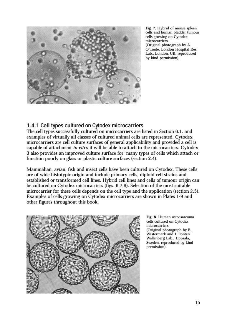
15 1.4.1 Cell types cultured on Cytodex microcarriers The cell types successfully cultured on microcarriers are listed in Section 6.1. and examples of virtually all classes of cultured animal cells are represented. Cytodex microcarriers are cell culture surfaces of general applicability and provided a cell is capable of attachment in vitro it will be able to attach to the microcarriers. Cytodex 3 also provides an improved culture surface for many types of cells which attach or function poorly on glass or plastic culture surfaces (section 2.4). Mammalian, avian, fish and insect cells have been cultured on Cytodex. These cells are of wide histotypic origin and include primary cells, diploid cell strains and established or transformed cell lines. Hybrid cell lines and cells of tumour origin can be cultured on Cytodex microcarriers (figs. 6,7,8). Selection of the most suitable microcarrier for these cells depends on the cell type and the application (section 2.5). Examples of cells growing on Cytodex microcarriers are shown in Plates 1-9 and other figures throughout this book. Fig. 7. Hybrid of mouse spleen cells and human bladder tumour cells growing on Cytodex microcarriers. (Original photograph by A. O’Toole, London Hospital Res. Lab., London, UK, reproduced by kind permission). Fig. 8. Human osteosarcoma cells cultured on Cytodex microcarriers. (Original photograph by B. Westermark and J. Pontén. Wallenberg Lab., Uppsala, Sweden, reproduced by kind permission)
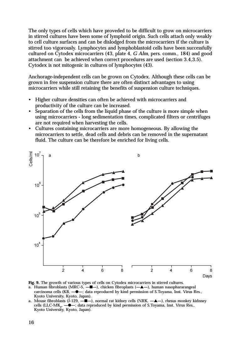
The only types of cells which have proveded to be difficult to grow on microcarriers in stirred cultures have been some of lymphoid origin.Such cells attach only weakly to cell culture surfaces and can be dislodged from the microcarriers if the culture is stirred too vigorously.Lymphocytes and lymphoblastoid cells have been successfully cultured on Cytodex microcarriers(43,plate 4.G Alm,pers.comm.184)and good attachment can be achieved when correct procedures are used(section 3.4,3.5). Cytodex is not mitogenic in cultures of lymphocytes(43). Anchorage-independent cells can be grown on Cytodex.Although these cells can be grown in free suspension culture there are often distinct advantages to using microcarriers while still retaining the benefits of suspension culture techniques. Higher culture densities can often be achieved with microcarriers and productivity of the culture can be increased. ane mone o e mr using microcarriers-long sedimentation b 105 10 10 246 a. sts -129, dTt Vmy Kyoto University.Kyoto.Japan). 16
16 The only types of cells which have proveded to be difficult to grow on microcarriers in stirred cultures have been some of lymphoid origin. Such cells attach only weakly to cell culture surfaces and can be dislodged from the microcarriers if the culture is stirred too vigorously. Lymphocytes and lymphoblastoid cells have been successfully cultured on Cytodex microcarriers (43, plate 4, G Alm, pers. comm., 184) and good attachment can be achieved when correct procedures are used (section 3.4,3.5). Cytodex is not mitogenic in cultures of lymphocytes (43). Anchorage-independent cells can be grown on Cytodex. Although these cells can be grown in free suspension culture there are often distinct advantages to using microcarriers while still retaining the benefits of suspension culture techniques. • Higher culture densities can often be achieved with microcarriers and productivity of the culture can be increased. • Separation of the cells from the liquid phase of the culture is more simple when using microcarriers - long sedimentation times, complicated filters or centrifuges are not required when harvesting the cells. • Cultures containing microcarriers are more homogeneous. By allowing the microcarriers to settle, dead cells and debris can be removed in the supernatant fluid. The culture can be therefore be enriched for living cells. Days 2 2 104 105 106 107 a b 4 6 8 8 4 6 Cells/ml Fig. 9. The growth of various types of cells on Cytodex microcarriers in stirred cultures. a. Human fibroblasts (MRC-5, —■—), chicken fibroplasts (—▲—), human nasophararangeal carcinoma cells (KB, —●—; data reproduced by kind permission of S.Toyama, Inst. Virus Res., Kyoto University, Kyoto, Japan). a. Mouse fibroblasts (J-129, —■—), normal rat kidney cells (NRK, —▲—), rhesus monkey kidnney cells (LLC-MK2 , —●—; data reproduced by kind permission of S.Toyama, Inst. Virus Res., Kyoto University, Kyoto, Japan)