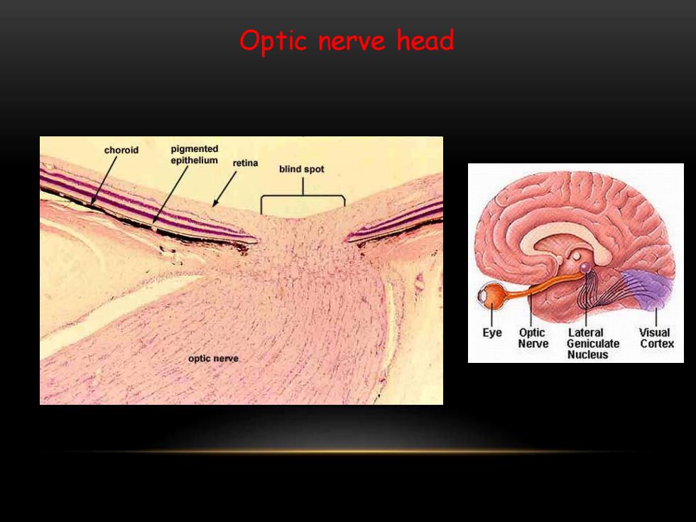
Optic nerve head choroid pigmented epithelium retina blind spot Eye Optic Lateral Visual Nerve Geniculate Cortex Nucleus
Optic nerve head
Outline Anatomy of the eye and the retina Retinal diagnostic test Diabetic Retinopathy (DR) Age-related Macular Degeneration (AMD)
• Anatomy of the eye and the retina • Retinal diagnostic test • Diabetic Retinopathy (DR) • Age-related Macular Degeneration (AMD) Outline

Retinal Diagnostic Tests ·Fundus Photography Fluorescein Angiography (FA) Optical Coherence Tomography (OCT) Ocular Ultrasonography Electroretinography (ERG)
• Fundus Photography • Fluorescein Angiography (FA) • Optical Coherence Tomography (OCT) • Ocular Ultrasonography • Electroretinography (ERG) Retinal Diagnostic Tests

Fundus Photography and Fluorescein Angiography (FA)
Fundus Photography and Fluorescein Angiography (FA)
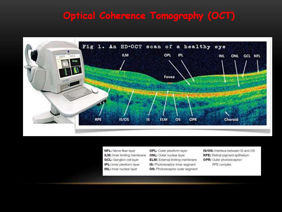
Optical Coherence Tomography (OCT) Fig 1.An ED-OCT scan of a healthy eye ILM OPL ONL GCL NFL Fovea NFL:Nerve fiber layer OPL:Outer plexitomm layer IS/OS:Interface between IS and OS ILM:Inner limiting membrane ONL:Outer nuclear layer RPE:Retinal pigment opithelum GCL:Gangion cell layer ELM:Externol limiting membrane IPL:inner plexifomm layer IS:Photoreceptor inner segment RPE complex
Optical Coherence Tomography (OCT)