4、荧光显微镜技术 ·原理:荧光物质,激发光,发射光 ·仪器:点光源(高压汞灯),滤色系统 荧光: 一自发荧光:如叶绿素、维生素A 诱发荧光:甲醛蒸汽处理诱发细胞和组织中 的生物单胺类产生炎光 一荧光染料染色荧光 ·应用
4、荧光显微镜技术 • 原理:荧光物质,激发光,发射光 • 仪器:点光源(高压汞灯),滤色系统 • 荧光: – 自发荧光:如叶绿素、维生素A – 诱发荧光:甲醛蒸汽处理诱发细胞和组织中 的生物单胺类产生荧光 – 荧光染料染色荧光 • 应用
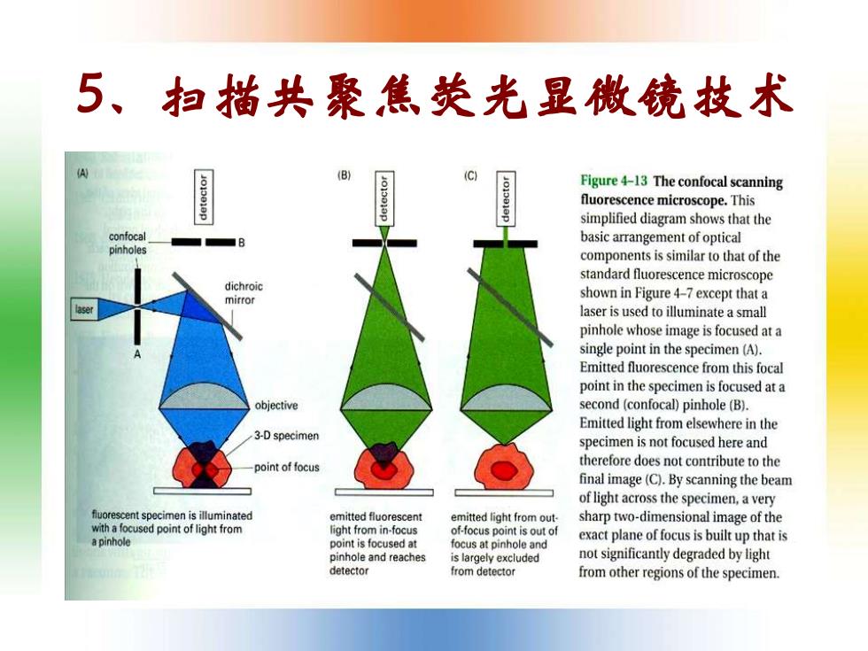
5、扫描共聚焦荧光显微镜技术 (B) Figure 4-13 The confocal scanning fluorescence microscope.This simplified diagram shows that the confocal basic arrangement of optical pinholes components is similar to that of the standard fluorescence microscope dichroic mirror shown in Figure 4-7 except that a laser is used to illuminate a small pinhole whose image is focused at a single point in the specimen (A). Emitted fluorescence from this focal point in the specimen is focused at a objective second (confocal)pinhole (B). Emitted light from elsewhere in the 3-D specimen specimen is not focused here and point of focus therefore does not contribute to the final image(C).By scanning the beam of light across the specimen,a very fluorescent specimen is illuminated emitted fluorescent emitted light from out. sharp two-dimensional image of the with a focused point of light from light from in-focus of-focus point is out of a pinhole point is focused at exact plane of focus is built up that is focus at pinhole and pinhole and reaches is largely excluded not significantly degraded by light detector from detector from other regions of the specimen
5、扫描共聚焦荧光显微镜技术
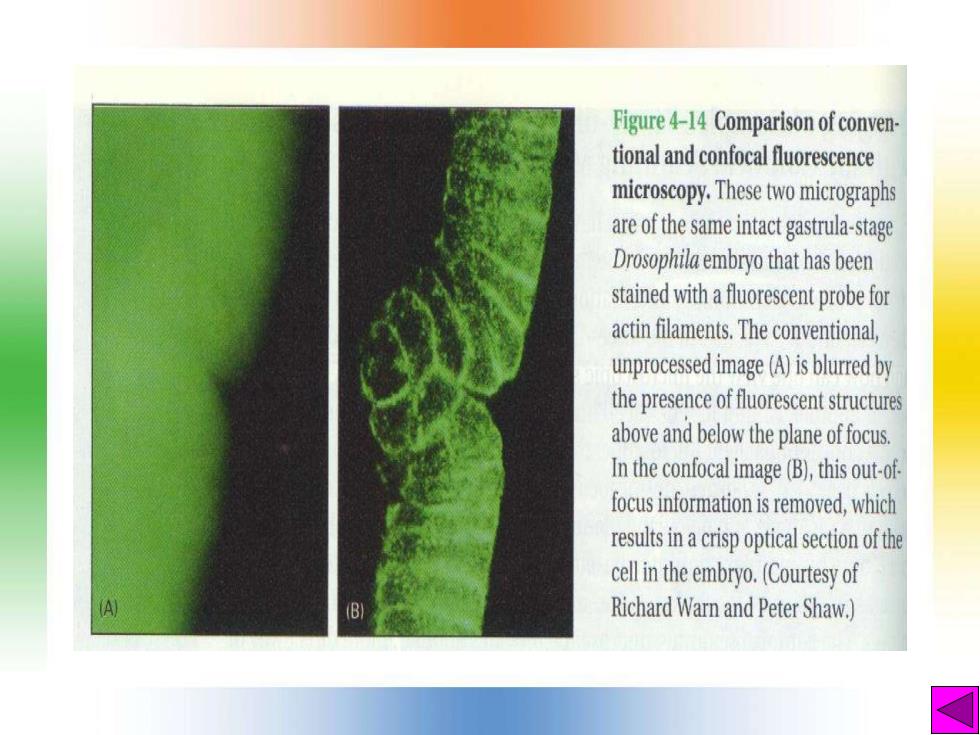
Figure 4-14 Comparison of conven- tional and confocal fluorescence microscopy.These two micrographs are of the same intact gastrula-stage Drosophila embryo that has been stained with a fuorescent probe for actin filaments.The conventional, unprocessed image (A)is blurred by the presence of fuoresetsructures above and below the plane of focus. In the confocaimage (B),this out-of focus information is removed,which resuiacrispopticlscofthe cell in the embryo.(Courtesy of (B) Richard Warn and Peter Shaw.)
(二)电子显微镜技术 ·电子显微镜的基本知识 一电镜与光镜的比较 一电子显微镜的基本构造 一电镜与光镜光路图的比较
(二)电子显微镜技术 • 电子显微镜的基本知识 – 电镜与光镜的比较 – 电子显微镜的基本构造 – 电镜与光镜光路图的比较
·主要电镜制片技术 一超薄切片技术 一冷冻独刻技术 一负染色技术 一电镜三维重构技术 。1 扫描电镜(SEM,Scanning electron microscope
• 主要电镜制片技术 – 超薄切片技术 – 冷冻蚀刻技术 – 负染色技术 – 电镜三维重构技术 • 扫描电镜(SEM,Scanning electron microscope)