microscopical features of tachizoites of Toxoplasma gondiiand peritoneal macrophages of mouse in peritoneal exudate(SEM)
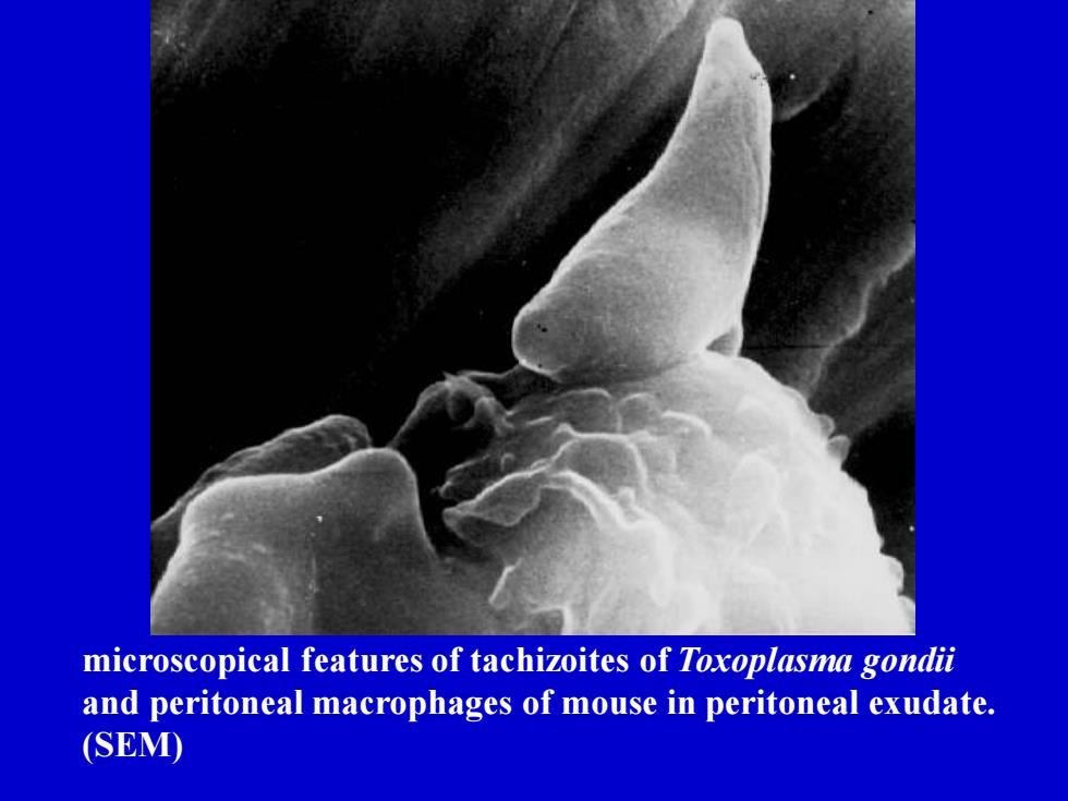
microscopical features of tachizoites of Toxoplasma gondii and peritoneal macrophages of mouse in peritoneal exudate. (SEM)

T. gondii: transmision electronmicroscopic pictureLongitudinal section of anendozoid
T. gondii: transmision electron microscopic picture. Longitudinal section of an endozoid
T. gondi: cross-section through an endozoid in an advanced stageof endodiogeny. The daugther cells appear to be surrounded.Ineach of these news cells there are two round bodies that lengthenformingthefirstrhoptries
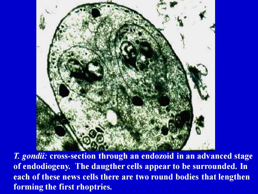
T. gondii: cross-section through an endozoid in an advanced stage of endodiogeny. The daugther cells appear to be surrounded. In each of these news cells there are two round bodies that lengthen forming the first rhoptries

Toxoplasmagondiiin tissuefroma cat
Toxoplasma gondii in tissue from a cat
toxoplasmic pseudocyst within an inflammatorytissue reaction(H&Estain)
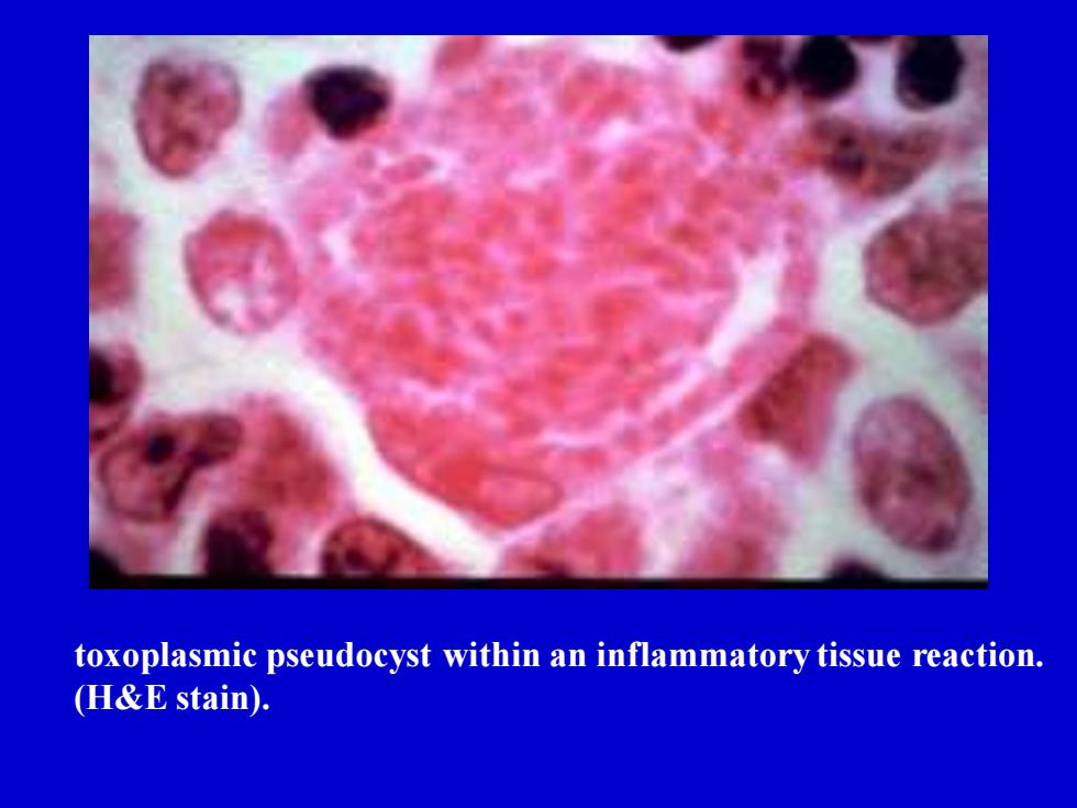
toxoplasmic pseudocyst within an inflammatory tissue reaction. (H&E stain)