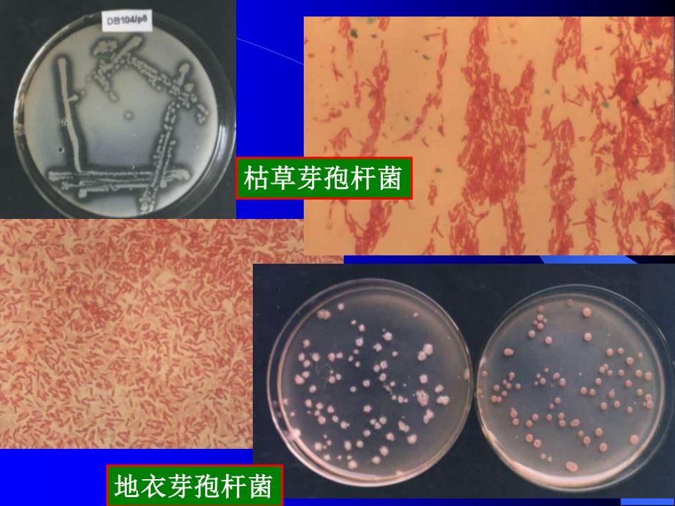
DB104/p6 枯草芽孢杆菌 地衣芽孢杆菌
枯草芽孢杆菌 地衣芽孢杆菌
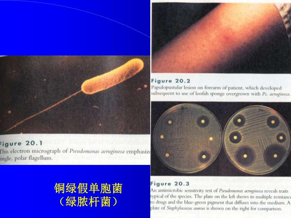
Figure 20.2 Papulopustular lesion on forearm of patient,which developed subsequent to use of loofah sponge overgrown with Ps.aenginos. igure 20.1 his electron micrograph of Pseudomonas aenginosa emphasiz ngle,polar flagellum. 铜绿假单胞菌 Figure 20.3 An antimicrobic sensitivity test of Pseudomonas aenginosa reveals traits (绿脓杆菌) ypical of the species.The plate on the left shows its multiple resistanc o drugs and the blue-green pigment that diffuses into the medium.A late of Staplryloccsres is shown on the right for comparison
铜绿假单胞菌 (绿脓杆菌)
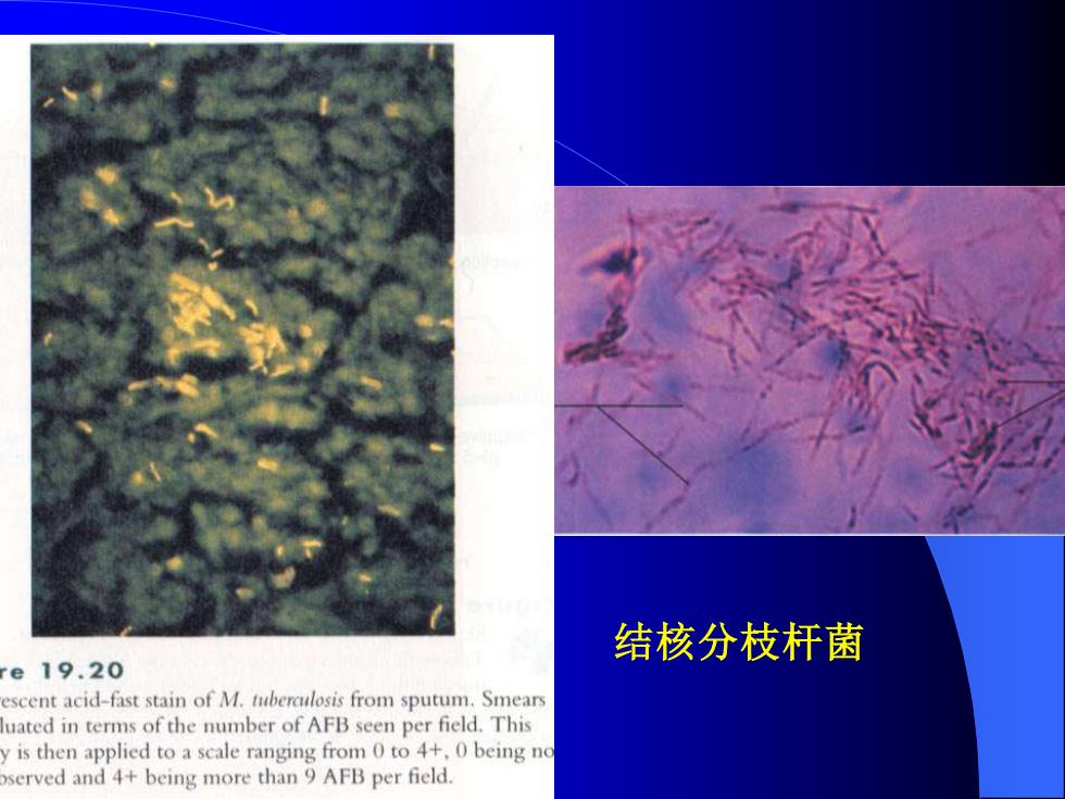
结核分枝杆菌 e19.20 escent acid-fast stain of M.tuberclosis from sputum.Smears uated in terms of the number of AFB seen per field.This is then applied to a scale ranging from 0 to 4+,0 being no served and 4+being more than 9 AFB per field
结核分枝杆菌
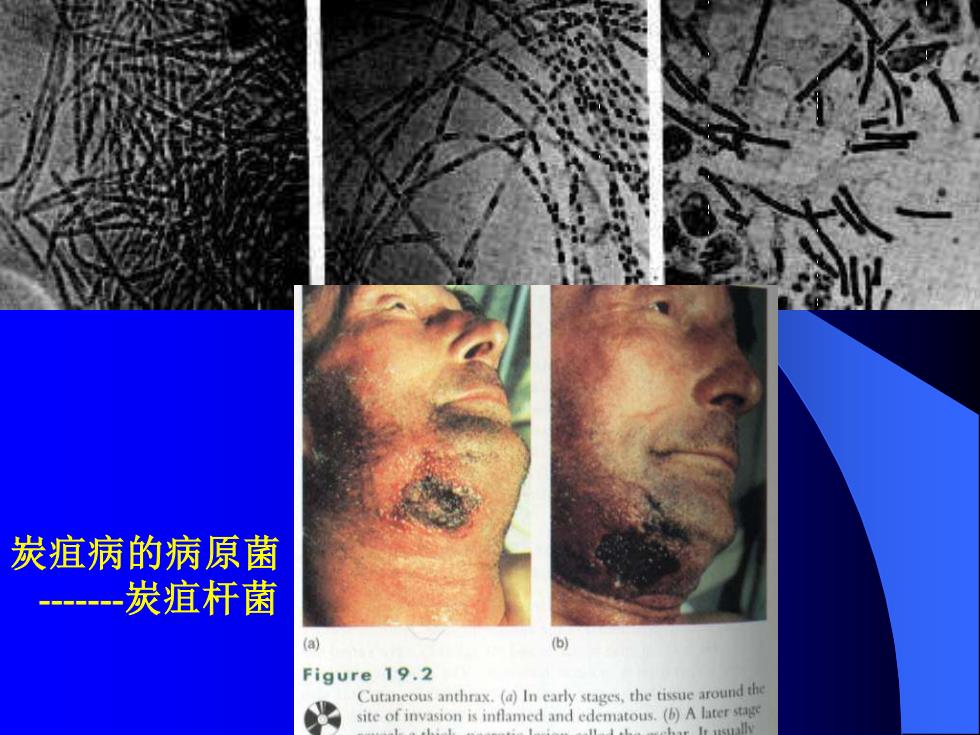
炭疽病的病原菌 -一炭疽杆菌 (a) b Figure 19.2 Cutaneous anthrax.(a)In early stages,the tissue around th iofomd andedematoA
炭疽病的病原菌 -炭疽杆菌
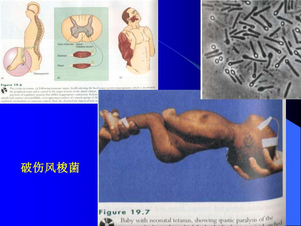
9ue9, 破伤风梭菌 Figure 19.7 Baby with neonatal tetanus.showing spastic paralysis of the
破伤风梭菌