axon may,however,branch into a set of collaterals allowing contact to be made with many other neurons.With respect to a particular neuron,other neurons that supply input are said to be afferent,while the given neuron's axonal output, regarded as a projection to other cells,is referred to as an efferent.Afferent axons are said to innervate a particular neuron and make contact with dendrites at the junctions called synapses.Here,the extremity of the axon,or axon terminal,comes into close proximity with a small part of the dendritic surface-the postsynaptic membrane.There is a gap,the synoptic cleft,between the presynaptic axon terminal membrane and its postsynaptic counterpart,which is of the order of 20 nanometres(2x10⁻⁸m)wide.Only a few synapses are shown in Figure 2.1 for the sake of clarity but the reader should imagine a profusion of these located over all dendrites and also,possibly,the cell body.The detailed synaptic structure is shown in schematic form as an inset in the figure. So much for neural structure;how does it support signal processing?At equilibrium,the neural membrane works to maintain an electrical imbalance of negatively and positively charged ions.These are atoms or molecules that have a surfeit or deficit of electrons,where each of the latter carries a single negative charge.The net result is that there is a potential difference across the membrane with the inside being negatively polarized by approximately 70mV with respect to the outside.Thus,if we could imagine applying a voltmeter to the membrane it would read 70mV,with the inside being more negative than the outside.The main point here is that a neural membrane can support electrical signals if its state of polarization or membrane potential is dynamically changed.To see this,consider the case of signal propagation along an axon as shown in Figure 2.2.Signals that are propagated along axons,or action potentials,all have the same characteristic shape,resembling sharp pulse-like spikes.Each graph shows a snapshot of the membrane potential along a segment of axon that is currently transmitting a single action potential,and the lower panel shows the situation at some later time with respect to the upper one.The ionic mechanisms at work to produce this process were first worked out by Hodgkin Huxley (1952).It relies 23
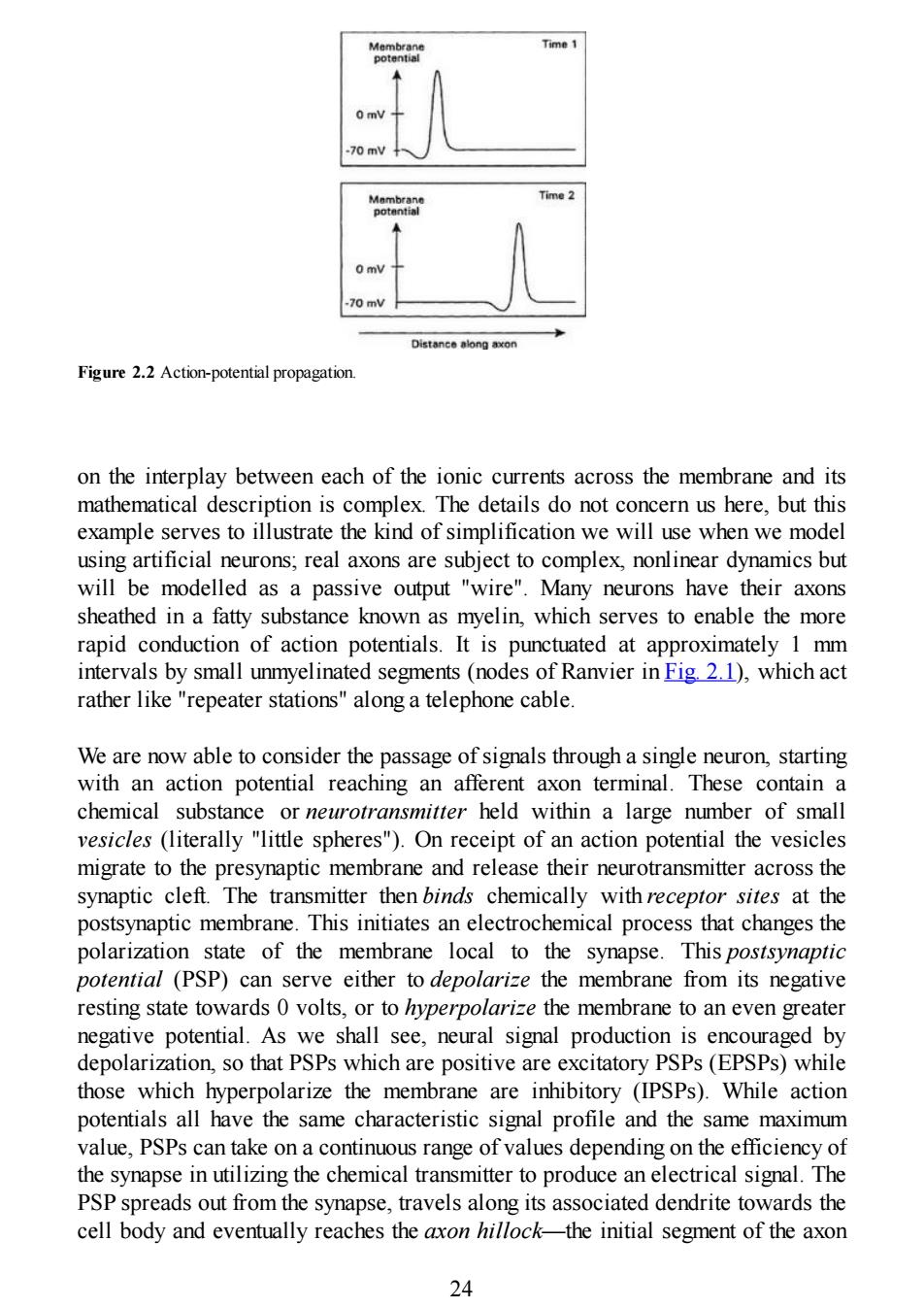
axon may, however, branch into a set of collaterals allowing contact to be made with many other neurons. With respect to a particular neuron, other neurons that supply input are said to be af erent, while the given neuron's axonal output, regarded as a projection to other cells, is referred to as an ef erent. Afferent axons are said to innervate a particular neuron and make contact with dendrites at the junctions called synapses. Here, the extremity of the axon, or axon terminal, comes into close proximity with a small part of the dendritic surface—the postsynaptic membrane. There is a gap, the synoptic cleft, between the presynaptic axon terminal membrane and its postsynaptic counterpart, which is of the order of 20 nanometres (2×10 -8m) wide. Only a few synapses are shown in Figure 2.1 for the sake of clarity but the reader should imagine a profusion of these located over all dendrites and also, possibly, the cell body. The detailed synaptic structure is shown in schematic form as an inset in the figure. So much for neural structure; how does it support signal processing? At equilibrium, the neural membrane works to maintain an electrical imbalance of negatively and positively charged ions. These are atoms or molecules that have a surfeit or deficit of electrons, where each of the latter carries a single negative charge. The net result is that there is a potential dif erence across the membrane with the inside being negatively polarized by approximately 70mV1 with respect to the outside. Thus, if we could imagine applying a voltmeter to the membrane it would read 70mV, with the inside being more negative than the outside. The main point here is that a neural membrane can support electrical signals if its state of polarization or membrane potential is dynamically changed. To see this, consider the case of signal propagation along an axon as shown in Figure 2.2. Signals that are propagated along axons, or action potentials, all have the same characteristic shape, resembling sharp pulse-like spikes. Each graph shows a snapshot of the membrane potential along a segment of axon that is currently transmitting a single action potential, and the lower panel shows the situation at some later time with respect to the upper one. The ionic mechanisms at work to produce this process were first worked out by Hodgkin & Huxley (1952). It relies 23
Time 1 70 mV Time 2 0 mV 70 mV Distance along axon Figure 2.2 Action-potential propagation on the interplay between each of the ionic currents across the membrane and its mathematical description is complex.The details do not concern us here,but this example serves to illustrate the kind of simplification we will use when we model using artificial neurons;real axons are subject to complex,nonlinear dynamics but will be modelled as a passive output "wire".Many neurons have their axons sheathed in a fatty substance known as myelin,which serves to enable the more rapid conduction of action potentials.It is punctuated at approximately 1 mm intervals by small unmyelinated segments (nodes of Ranvier in Fig.2.1),which act rather like "repeater stations"along a telephone cable. We are now able to consider the passage of signals through a single neuron,starting with an action potential reaching an afferent axon terminal.These contain a chemical substance or neurotransmitter held within a large number of small vesicles (literally "little spheres").On receipt of an action potential the vesicles migrate to the presynaptic membrane and release their neurotransmitter across the synaptic cleft.The transmitter then binds chemically with receptor sites at the postsynaptic membrane.This initiates an electrochemical process that changes the polarization state of the membrane local to the synapse.This postsynaptic potential (PSP)can serve either to depolarize the membrane from its negative resting state towards 0 volts,or to hyperpolarize the membrane to an even greater negative potential.As we shall see,neural signal production is encouraged by depolarization,so that PSPs which are positive are excitatory PSPs(EPSPs)while those which hyperpolarize the membrane are inhibitory (IPSPs).While action potentials all have the same characteristic signal profile and the same maximum value,PSPs can take on a continuous range of values depending on the efficiency of the synapse in utilizing the chemical transmitter to produce an electrical signal.The PSP spreads out from the synapse,travels along its associated dendrite towards the cell body and eventually reaches the axon hillock-the initial segment of the axon 24
Figure 2.2 Action-potential propagation. on the interplay between each of the ionic currents across the membrane and its mathematical description is complex. The details do not concern us here, but this example serves to illustrate the kind of simplification we will use when we model using artificial neurons; real axons are subject to complex, nonlinear dynamics but will be modelled as a passive output "wire". Many neurons have their axons sheathed in a fatty substance known as myelin, which serves to enable the more rapid conduction of action potentials. It is punctuated at approximately 1 mm intervals by small unmyelinated segments (nodes of Ranvier in Fig. 2.1), which act rather like "repeater stations" along a telephone cable. We are now able to consider the passage of signals through a single neuron, starting with an action potential reaching an afferent axon terminal. These contain a chemical substance or neurotransmitter held within a large number of small vesicles (literally "little spheres"). On receipt of an action potential the vesicles migrate to the presynaptic membrane and release their neurotransmitter across the synaptic cleft. The transmitter then binds chemically with receptor sites at the postsynaptic membrane. This initiates an electrochemical process that changes the polarization state of the membrane local to the synapse. This postsynaptic potential (PSP) can serve either to depolarize the membrane from its negative resting state towards 0 volts, or to hyperpolarize the membrane to an even greater negative potential. As we shall see, neural signal production is encouraged by depolarization, so that PSPs which are positive are excitatory PSPs (EPSPs) while those which hyperpolarize the membrane are inhibitory (IPSPs). While action potentials all have the same characteristic signal profile and the same maximum value, PSPs can take on a continuous range of values depending on the efficiency of the synapse in utilizing the chemical transmitter to produce an electrical signal. The PSP spreads out from the synapse, travels along its associated dendrite towards the cell body and eventually reaches the axon hillock—the initial segment of the axon 24
where it joins the soma.Concurrent with this are thousands of other synaptic events distributed over the neuron.These result in a plethora of PSPs,which are continually arriving at the axon hillock where they are summed together to produce a resultant membrane potential. Each contributory PSP at the axon hillock exists for an extended time (order of milliseconds)before it eventually decays so that,if two PSPs arrive slightly out of synchrony,they may still interact in the summation process.On the other hand, suppose two synaptic events take place with one close to and another remote from the soma,by virtue of being at the end of a long dendritic branch.By the time the PSP from the distal (remote)synapse has reached the axon hillock,that originating close to the soma will have decayed.Thus,although the initiation of PSPs may take place in synchrony,they may not be effective in combining to generate action potentials.It is apparent,therefore,that a neuron sums or integrates its PSPs over both space and time.Substantial modelling effort-much of it pioneered by Rail (1957,1959)-has gone into describing the conduction of PSPs along dendrites and their subsequent interaction although,as in the case of axons,connectionist models usually treat these as passive wires with no temporal characteristics. The integrated PSP at the axon hillock will affect its membrane potential and,if this exceeds a certain threshold (typically about -50mV),an action potential is generated,which then propagates down the axon,along any collaterals,eventually reaching axon terminals resulting in a shower of synaptic events at neighbouring neurons "downstream"of our original cell.In reality the "threshold"is an emergent or meta-phenomenon resulting from the nonlinear nature of the Hodgkin-Huxley dynamics and,under certain conditions,it can be made to change.However,for many purposes it serves as a suitable high-level description of what actually occurs.After an action potential has been produced,the ionic metabolites used in its production have been depleted and there is a short refractory period during which,no matter what value the membrane potential takes,there can be no initiation of another action potential. It is useful at this stage to summarize what we have learnt so far about the functionality of real neurons with an eye to the simplification required for modelling their artificial counterparts. - Signals are transmitted between neurons by action potentials,which have a stereotypical profile and display an "all-or-nothing"character;there is no such thing as halfan action potential. -When an action potential impinges on a neuronal input(synapse)the effect is a PSP,which is variable or graded and depends on the physicochemical properties of the synapse. 25
where it joins the soma. Concurrent with this are thousands of other synaptic events distributed over the neuron. These result in a plethora of PSPs, which are continually arriving at the axon hillock where they are summed together to produce a resultant membrane potential. Each contributory PSP at the axon hillock exists for an extended time (order of milliseconds) before it eventually decays so that, if two PSPs arrive slightly out of synchrony, they may still interact in the summation process. On the other hand, suppose two synaptic events take place with one close to and another remote from the soma, by virtue of being at the end of a long dendritic branch. By the time the PSP from the distal (remote) synapse has reached the axon hillock, that originating close to the soma will have decayed. Thus, although the initiation of PSPs may take place in synchrony, they may not be effective in combining to generate action potentials. It is apparent, therefore, that a neuron sums or integrates its PSPs over both space and time. Substantial modelling effort—much of it pioneered by Rail (1957, 1959)—has gone into describing the conduction of PSPs along dendrites and their subsequent interaction although, as in the case of axons, connectionist models usually treat these as passive wires with no temporal characteristics. The integrated PSP at the axon hillock will affect its membrane potential and, if this exceeds a certain threshold (typically about -50mV), an action potential is generated, which then propagates down the axon, along any collaterals, eventually reaching axon terminals resulting in a shower of synaptic events at neighbouring neurons "downstream" of our original cell. In reality the "threshold" is an emergent or meta-phenomenon resulting from the nonlinear nature of the Hodgkin-Huxley dynamics and, under certain conditions, it can be made to change. However, for many purposes it serves as a suitable high-level description of what actually occurs. After an action potential has been produced, the ionic metabolites used in its production have been depleted and there is a short refractory period during which, no matter what value the membrane potential takes, there can be no initiation of another action potential. It is useful at this stage to summarize what we have learnt so far about the functionality of real neurons with an eye to the simplification required for modelling their artificial counterparts. – Signals are transmitted between neurons by action potentials, which have a stereotypical profile and display an "all-or-nothing" character; there is no such thing as half an action potential. – When an action potential impinges on a neuronal input (synapse) the effect is a PSP, which is variable or graded and depends on the physicochemical properties of the synapse. 25
The PSPs may be excitatory or inhibitory -The PSPs are summed together at the axon hillock with the result expressed as its membrane potential. -If this potential exceeds a threshold an action potential is initiated that proceeds along the axon. Several things have been deliberately omitted here.First,the effect that synaptic structure can have on the value of the PSP.Factors that may play a role here include the type and availability of neurotransmitter,the postsynaptic receptors and synaptic geometry.Secondly,the spatio-temporal interdependencies of PSPs resulting from dendritic geometry whereby,for example,synapses that are remote from each other may not effectively combine.Finally,we have said nothing about t he dynamics of action-potential generation and propagation.However,our summary will serve as a point of departure for defining the kind of artificial neurons described in this book.More biologically realistic models rely on solving Hodgkin-Huxley-type dynamics and modelling dendrites at the electrical circuit level;details of these methods can be found in the review compilation of Koch Segev (1989). 2.1.1 Glossary of terms Those terms in italics may be cross-referenced in this glossary action potential The stereotypical voltage spike that constitutes an active output from a neuron.They are propagated along the axon to other neurons. afferent With respect to a particular neuron,an axon that impinges on (or innervates)that neuron. arbor Usually used in the context of a dendritic arbor-the tree-like structure associated with dendritic branching. axon The fibre that emanates from the neuron cell body or soma and that conducts action potentials to other neurons. axon hillock The junction of the axon and cell body or soma.The place where action potentials are initiated if the membrane potential exceeds a threshold. axon terminal An axon may branch into several collaterals,each terminating at an 26
– The PSPs may be excitatory or inhibitory. – The PSPs are summed together at the axon hillock with the result expressed as its membrane potential. – If this potential exceeds a threshold an action potential is initiated that proceeds along the axon. Several things have been deliberately omitted here. First, the effect that synaptic structure can have on the value of the PSP. Factors that may play a role here include the type and availability of neurotransmitter, the postsynaptic receptors and synaptic geometry. Secondly, the spatio-temporal interdependencies of PSPs resulting from dendritic geometry whereby, for example, synapses that are remote from each other may not effectively combine. Finally, we have said nothing about t h e dynamics of action-potential generation and propagation. However, our summary will serve as a point of departure for defining the kind of artificial neurons described in this book. More biologically realistic models rely on solving Hodgkin-Huxley-type dynamics and modelling dendrites at the electrical circuit level; details of these methods can be found in the review compilation of Koch & Segev (1989). 2.1.1 Glossary of terms Those terms in italics may be cross-referenced in this glossary. action potential The stereotypical voltage spike that constitutes an active output from a neuron. They are propagated along the axon to other neurons. afferent With respect to a particular neuron, an axon that impinges on (or innervates) that neuron. arbor Usually used in the context of a dendritic arbor—the tree-like structure associated with dendritic branching. axon The fibre that emanates from the neuron cell body or soma and that conducts action potentials to other neurons. axon hillock The junction of the axon and cell body or soma. The place where action potentials are initiated if the membrane potential exceeds a threshold. axon terminal An axon may branch into several collaterals, each terminating at an 26

axon terminal,which constitutes the presynaptic component of a synapse chemical binding The process in which a neurotransmitter joins chemically with a receptor site thereby initiating a PSP. collateral An axon may divide into many collateral branches allowing contact with many other neurons or many contacts with one neuron. dendrite One of the branching fibres of a neuron,which convey input information via PSPs. de polarization The membrane potential of the neuron has a negative resting or equilibrium value.Making this less negative leads to a depolarization.Sufficient depolarization at the axon hillock will give rise to an action potential. efferent A neuron sends efferent axon collaterals to other neurons. EPSP Excitatory Postsynaptic Potential.A PSP that acts to depolarize the neural membrane hyperpolarization The membrane potential of the neuron has a negative resting or equilibrium value.Making this more negative leads to a hyperpolarization and inhibits the action of EPSPs,which are trying to depolarize the membrane. innervate Neuron 4 sending signals to neuron B is said to innervate neuron B. IPSP Inhibitory Postsynaptic Potential.A PSP that acts to hyperpolarize the neural membrane. membrane potential The voltage difference at any point across the neural membrane. neurotransmitter The chemical substance that mediates synaptic activity by propagation across the synaptic cleft. organelle Subcellular components that partake in metabolism,etc. postsynaptic membrane That part of a synapse which is located on the dendrite and consists of the dendritic membrane together with receptor sites. potential difference The voltage difference across the cell membrane. presynaptic membrane That part of a synapse which is located on the axon 27
axon terminal, which constitutes the presynaptic component of a synapse. chemical binding The process in which a neurotransmitter joins chemically with a receptor site thereby initiating a PSP. collateral An axon may divide into many collateral branches allowing contact with many other neurons or many contacts with one neuron. dendrite One of the branching fibres of a neuron, which convey input information via PSPs. depolarization The membrane potential of the neuron has a negative resting or equilibrium value. Making this less negative leads to a depolarization. Sufficient depolarization at the axon hillock will give rise to an action potential. efferent A neuron sends efferent axon collaterals to other neurons. EPSP Excitatory Postsynaptic Potential. A PSP that acts to depolarize the neural membrane. hyperpolarization The membrane potential of the neuron has a negative resting or equilibrium value. Making this more negative leads to a hyperpolarization and inhibits the action of EPSPs, which are trying to depolarize the membrane. innervate Neuron A sending signals to neuron B is said to innervate neuron B. IPSP Inhibitory Postsynaptic Potential. A PSP that acts to hyperpolarize the neural membrane. membrane potential The voltage difference at any point across the neural membrane. neurotransmitter The chemical substance that mediates synaptic activity by propagation across the synaptic cleft. organelle Subcellular components that partake in metabolism, etc. postsynaptic membrane That part of a synapse which is located on the dendrite and consists of the dendritic membrane together with receptor sites. potential difference The voltage difference across the cell membrane. presynaptic membrane That part of a synapse which is located on the axon 27